

Links between alpha-catenin, NF-kappaB, and squamous cell carcinoma in skin
- PMID: 16452166
- PMCID: PMC1413714
- DOI: 10.1073/pnas.0510422103
Links between alpha-catenin, NF-kappaB, and squamous cell carcinoma in skin
Abstract
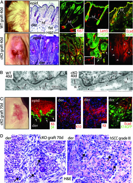
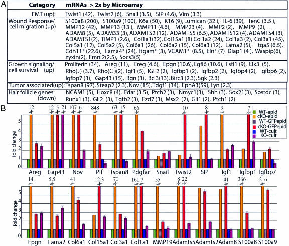
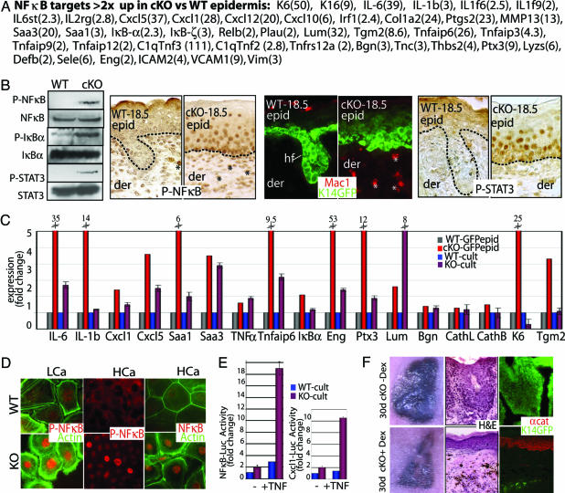
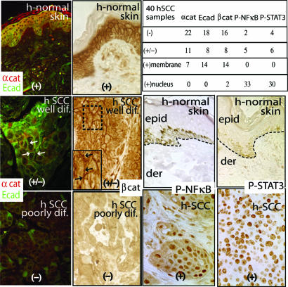
Cancers display a diverse set of cellular defects, which are thought to be elicited by multiple genetic mutations. In this study, we show that when a single adherens junction protein, alpha-catenin, is removed by conditional targeting, the entire skin epidermis systematically transforms to a hyperproliferative, invasive tissue replete with inflammation. Transcriptional profiling and biochemical analyses reveal that alpha-catenin ablation is accompanied by activation of NF-kappaB and its proinflammatory target genes, along with genes involved in proliferation, wound healing, angiogenesis, and metastasis. Many of these alterations occur in vitro and in the embryo, and thus seem at least partly to be intrinsic to the loss of alpha-catenin. We show that reductions in alpha-catenin, activation of NF-kappaB, and inflammation are common features of human squamous cell carcinomas of the skin.
Conflict of interest statement
Conflict of interest statement: No conflicts declared.
Figures
Similar articles
-
Nuclear factor-kappaB is an important modulator of the altered gene expression profile and malignant phenotype in squamous cell carcinoma.Cancer Res. 2004 Sep 15;64(18):6511-23. doi: 10.1158/0008-5472.CAN-04-0852. Cancer Res. 2004. PMID: 15374962
-
Molecular profiling of transformed and metastatic murine squamous carcinoma cells by differential display and cDNA microarray reveals altered expression of multiple genes related to growth, apoptosis, angiogenesis, and the NF-kappaB signal pathway.Cancer Res. 2001 Jun 15;61(12):4797-808. Cancer Res. 2001. PMID: 11406555
-
Epidermal overexpression of transgenic ΔNp63 promotes type 2 immune and myeloid inflammatory responses and hyperplasia via NF-κB activation.J Pathol. 2014 Feb;232(3):356-68. doi: 10.1002/path.4302. J Pathol. 2014. PMID: 24258200
-
NF-kappaB gene signatures and p53 mutations in head and neck squamous cell carcinoma.Clin Cancer Res. 2007 Oct 1;13(19):5663-4. doi: 10.1158/1078-0432.CCR-07-1544. Clin Cancer Res. 2007. PMID: 17908953 Review. No abstract available.
-
Role of activated nuclear factor-kappaB in the pathogenesis and therapy of squamous cell carcinoma of the head and neck.Head Neck. 2007 Oct;29(10):959-71. doi: 10.1002/hed.20615. Head Neck. 2007. PMID: 17405170 Review.
Cited by
-
Nuclear signaling from cadherin adhesion complexes.Curr Top Dev Biol. 2015;112:129-96. doi: 10.1016/bs.ctdb.2014.11.018. Epub 2015 Feb 12. Curr Top Dev Biol. 2015. PMID: 25733140 Free PMC article. Review.
-
The role of barrier genes in epidermal malignancy.Oncogene. 2016 Nov 3;35(44):5705-5712. doi: 10.1038/onc.2016.84. Epub 2016 Apr 4. Oncogene. 2016. PMID: 27041586 Review.
-
N-cadherin-mediated cell adhesion restricts cell proliferation in the dorsal neural tube.Mol Biol Cell. 2011 May;22(9):1505-15. doi: 10.1091/mbc.E10-08-0675. Epub 2011 Mar 9. Mol Biol Cell. 2011. PMID: 21389116 Free PMC article.
-
Dissecting the role of cadherin-catenin proteins in mammalian epidermis.Proc Natl Acad Sci U S A. 2008 Oct 7;105(40):15225-6. doi: 10.1073/pnas.0808458105. Epub 2008 Oct 1. Proc Natl Acad Sci U S A. 2008. PMID: 18832145 Free PMC article. No abstract available.
-
α(E)-catenin regulates BMP-7 expression and migration in renal epithelial cells.Am J Nephrol. 2014;39(5):409-17. doi: 10.1159/000362250. Epub 2014 May 6. Am J Nephrol. 2014. PMID: 24818804 Free PMC article.
References
Publication types
MeSH terms
Substances
Grants and funding
LinkOut - more resources
Full Text Sources
Medical
Molecular Biology Databases
