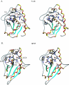
Solution structure of a low-molecular-weight protein tyrosine phosphatase from Bacillus subtilis
- PMID: 16452434
- PMCID: PMC1367216
- DOI: 10.1128/JB.188.4.1509-1517.2006
Solution structure of a low-molecular-weight protein tyrosine phosphatase from Bacillus subtilis
Abstract
The low-molecular-weight (LMW) protein tyrosine phosphatases (PTPs) exist ubiquitously in prokaryotes and eukaryotes and play important roles in cellular processes. We report here the solution structure of YwlE, an LMW PTP identified from the gram-positive bacteria Bacillus subtilis. YwlE consists of a twisted central four-stranded parallel beta-sheet with seven alpha-helices packing on both sides. Similar to LMW PTPs from other organisms, the conformation of the YwlE active site is favorable for phosphotyrosine binding, indicating that it may share a common catalytic mechanism in the hydrolysis of phosphate on tyrosine residue in proteins. Though the overall structure resembles that of the eukaryotic LMW PTPs, significant differences were observed around the active site. Residue Asp115 is likely interacting with residue Arg13 through electrostatic interaction or hydrogen bond interaction to stabilize the conformation of the active cavity, which may be a unique character of bacterial LMW PTPs. Residues in the loop region from Phe40 to Thr48 forming a wall of the active cavity are more flexible than those in other regions. Ala41 and Gly45 are located near the active cavity and form a noncharged surface around it. These unique properties demonstrate that this loop may be involved in interaction with specific substrates. In addition, the results from spin relaxation experiments elucidate further insights into the mobility of the active site. The solution structure in combination with the backbone dynamics provides insights into the mechanism of substrate specificity of bacterial LMW PTPs.
Figures


Similar articles
-
Three-dimensional structure and ligand interactions of the low molecular weight protein tyrosine phosphatase from Campylobacter jejuni.Protein Sci. 2006 Oct;15(10):2381-94. doi: 10.1110/ps.062279806. Protein Sci. 2006. PMID: 17008719 Free PMC article.
-
In vitro characterization of the Bacillus subtilis protein tyrosine phosphatase YwqE.J Bacteriol. 2005 May;187(10):3384-90. doi: 10.1128/JB.187.10.3384-3390.2005. J Bacteriol. 2005. PMID: 15866923 Free PMC article.
-
Mechanistic studies on protein tyrosine phosphatases.Prog Nucleic Acid Res Mol Biol. 2003;73:171-220. doi: 10.1016/s0079-6603(03)01006-7. Prog Nucleic Acid Res Mol Biol. 2003. PMID: 12882518 Review.
-
The solution structure of Escherichia coli Wzb reveals a novel substrate recognition mechanism of prokaryotic low molecular weight protein-tyrosine phosphatases.J Biol Chem. 2006 Jul 14;281(28):19570-7. doi: 10.1074/jbc.M601263200. Epub 2006 May 1. J Biol Chem. 2006. PMID: 16651264
-
Low molecular weight protein tyrosine phosphatase: Multifaceted functions of an evolutionarily conserved enzyme.Biochim Biophys Acta. 2016 Oct;1864(10):1339-55. doi: 10.1016/j.bbapap.2016.07.001. Epub 2016 Jul 13. Biochim Biophys Acta. 2016. PMID: 27421795 Review.
Cited by
-
Crystal structures of Wzb of Escherichia coli and CpsB of Streptococcus pneumoniae, representatives of two families of tyrosine phosphatases that regulate capsule assembly.J Mol Biol. 2009 Sep 25;392(3):678-88. doi: 10.1016/j.jmb.2009.07.026. Epub 2009 Jul 16. J Mol Biol. 2009. PMID: 19616007 Free PMC article.
-
Unique structural characteristics of the rabbit prion protein.J Biol Chem. 2010 Oct 8;285(41):31682-93. doi: 10.1074/jbc.M110.118844. Epub 2010 Jul 16. J Biol Chem. 2010. PMID: 20639199 Free PMC article.
-
Three-dimensional structure and ligand interactions of the low molecular weight protein tyrosine phosphatase from Campylobacter jejuni.Protein Sci. 2006 Oct;15(10):2381-94. doi: 10.1110/ps.062279806. Protein Sci. 2006. PMID: 17008719 Free PMC article.
-
Protein-tyrosine phosphorylation in Bacillus subtilis: a 10-year retrospective.Front Microbiol. 2015 Jan 23;6:18. doi: 10.3389/fmicb.2015.00018. eCollection 2015. Front Microbiol. 2015. PMID: 25667587 Free PMC article.
-
The role of bacterial protein tyrosine phosphatases in the regulation of the biosynthesis of secreted polysaccharides.Antioxid Redox Signal. 2014 May 10;20(14):2274-89. doi: 10.1089/ars.2013.5726. Epub 2014 Mar 11. Antioxid Redox Signal. 2014. PMID: 24295407 Free PMC article. Review.
References
-
- Berti, A., S. Rigacci, G. Raugei, D. Degl'Innocenti, and G. Ramponi. 1994. Inhibition of cellular response to platelet-derived growth factor by low M(r) phosphotyrosine protein phosphatase overexpression. FEBS Lett. 349:7-12. - PubMed
-
- Case, D. A., D. A. Pearlman, J. W. Caldwell, T. E. Cheatham III, J. Wang, W. S. Ross, C. L. Simmerling, T. A. Darden, K. M. Merz, R. V. Stanton, A. L. Cheng, J. J. Vincent, M. Crowley, V. Tsui, H. Gohlke, R. J. Radmer, Y. Duan, J. Pitera, I. Massova, G. L. Seibel, U. C. Singh, P. K. Weiner, and P. A. Kollman. 2002. AMBER 7, p. 130-131. University of California, San Francisco, Calif.
-
- Chiarugi, P., M. L. Taddei, P. Cirri, D. Talini, F. Buricchi, G. Camici, G. Manao, G. Raugei, and G. Ramponi. 2000. Low molecular weight protein-tyrosine phosphatase controls the rate and the strength of NIH-3T3 cells adhesion through its phosphorylation on tyrosine 131 or 132. J. Biol. Chem. 275:37619-37627. - PubMed
-
- Chiarugi, P., P. Cirri, G. Raugei, G. Camici, F. Dolfi, A. Berti, and G. Ramponi. 1995. PDGF receptor as a specific in vivo target for low Mr phosphotyrosine protein phosphatase. FEBS Lett. 372:49-53. - PubMed
-
- Chiarugi, P., P. Cirri, M. L. Taddei, E. Giannoni, G. Camici, G. Manao, G. Raugei, and G. Ramponi. 2000. The low M(r) protein-tyrosine phosphatase is involved in Rho-mediated cytoskeleton rearrangement after integrin and platelet-derived growth factor stimulation. J. Biol. Chem. 275:4640-4646. - PubMed
Publication types
MeSH terms
Substances
Associated data
- Actions
LinkOut - more resources
Full Text Sources

