. 2006 Feb;188(4):1534-9.
doi: 10.1128/JB.188.4.1534-1539.2006.

In the Bacillus stearothermophilus DnaB-DnaG complex, the activities of the two proteins are modulated by distinct but overlapping networks of residues

Affiliations

In the Bacillus stearothermophilus DnaB-DnaG complex, the activities of the two proteins are modulated by distinct but overlapping networks of residues

Jenny Thirlway et al. J Bacteriol. 2006 Feb.

Abstract

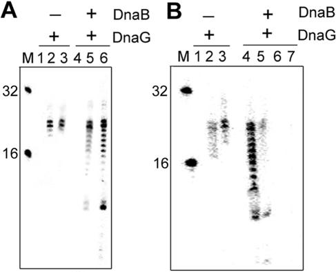
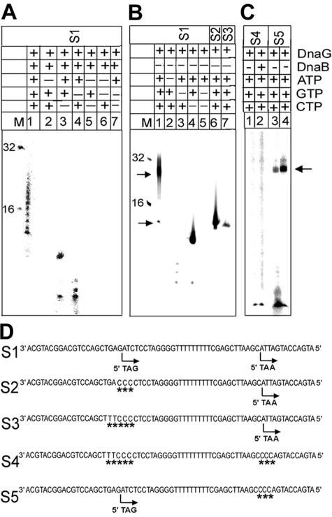
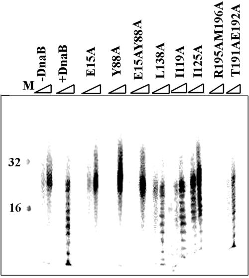
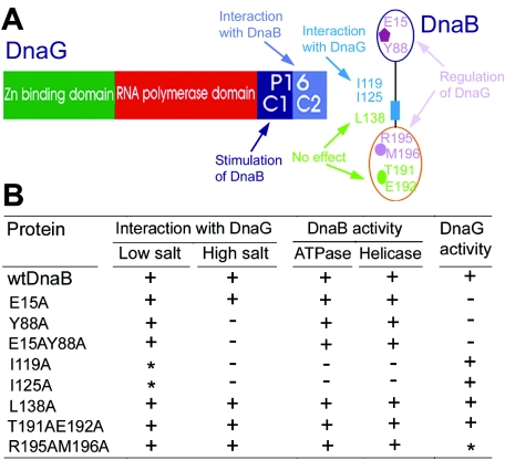
We demonstrate the primase activity of Bacillus stearothermophilus DnaG and show that it initiates at 3'-ATC-5' and 3'-ATT-5' sites synthesizing primers that are 22 or 23 nucleotides long. In the presence of the helicase DnaB the size distribution of primers is different, and a range of additional smaller primers are also synthesized. Nine residues from the N- and C-terminal domains of DnaB, as well as its linker region, have been reported previously to affect this interaction. In Bacillus stearothermophilus only three residues from the linker region (I119 and I125) and the N-terminal domain (Y88) of DnaB have been shown previously to have direct structural importance, and I119 and I125 mediate DnaG-induced effects on DnaB activity. The functions of the other residues (L138, T191, E192, R195, and M196) are still a mystery. Here we show that the E15A, Y88A, and E15A Y88A mutants bind DnaG but are not able to modulate primer size, whereas the R195A M196A mutant inhibited the primase activity. Therefore, four of these residues, E15 and Y88 (N-terminal domain) and R195 and M196 (C-terminal domain), mediate DnaB-induced effects on DnaG activity. Overall, the data suggest that the effects of DnaB on DnaG activity and vice versa are mediated by distinct but overlapping networks of residues.


Figures

FIG. 1.
FIG. 1.
General priming activity of DnaG. (A) General priming assays with substrate S1 and DnaG or DnaG plus DnaB, as indicated at the top. Reactions were carried out for 10, 30, and 40 min (for DnaG, lanes 1 to 3, respectively; for DnaG plus DnaB, lanes 4 to 6, respectively) at 50°C. DnaB modulates the activity of DnaG. In the presence of DnaB the distribution of primers synthesized by DnaG is different. In lanes 1 to 3, primers that are 22 or 23 nucleotides long are visible, whereas in lanes 4 to 6 a range of additional smaller primers are visible. (B) The relative molar concentrations of the two enzymes are critical for maximal modulation of the priming activity. Lanes 1 to 3 (10, 20, and 40 min, respectively) show a time course of the priming reaction in the absence of DnaB. Lanes 4 to 7 show the 40-min results for reactions performed in the presence of various concentrations of DnaB (lane 4, 2 nM; lane 5, 0.2 nM; lane 6, 20 nM; lane 7, 60 nM). All reactions were carried out with oligonucleotide substrate S1 at 50°C and 360 nM DnaG. Lane M contained oligonucleotide size markers.
FIG. 2.
FIG. 2.
General priming assays with substrate S1 and depleted combinations of nucleotides, as indicated at the top. Reactions were carried out as described in Materials and Methods in the presence of 360 nM DnaG for 60 min at 50°C, either in the presence (A) or in the absence (B) of 2 nM DnaB. Lanes 6 and 7 in panel B show reactions with oligonucleotide substrates S2 and S3, respectively. The arrows in lane 1 in panel B indicate primers initiating from the 5′-ATC-3′ (top arrow) and 5′-ATT-3′ (bottom arrow) sites. (C) Priming reactions with substrates S4 and S5. Most reactions were carried out for 60 min at 50°C with 360 nM DnaG in the absence of DnaB. Lane 2 shows the results of a reaction performed in the presence of 2 nM DnaB. The reaction with substrate S4 gave no primers (lanes 1 and 2). Lane M contained oligonucleotide size markers. (D) All the oligonucleotide substrates used in this study. Initiation sites are indicated by arrows, and sequence differences compared to substrate S1 are indicated by asterisks.
FIG. 3.
FIG. 3.
Effects of DnaB mutants on the activity of DnaG. Time course priming reactions (10, 20, and 40 min) were performed with substrate S1 in the presence of 2 nM DnaB mutants, as indicated at the top. Control reactions in the presence and absence of wild-type DnaB are indicated by +DnaB and −DnaB, respectively. Lane M contained size markers.
FIG. 4.
FIG. 4.
(A) Schematic representation of the domain organization of DnaB and DnaG and summary of the interaction interface between the two proteins. DnaG consists of three domains, a Zn-binding N-terminal domain (14), a central polymerization domain (9), and the C-terminal P16 domain that interacts with DnaB (13, 16, 19). The P16 domain consists of two subdomains, Subdomains C1 and C2. C2 structurally mediates the interaction with DnaB, while C1 mediates the functional stimulation of DnaB (16, 19). DnaB consists of N- and C-terminal domains (2, 9, 22). Residues from the flexible hinge region that connects the two domains are involved directly in the interaction (20), whereas the roles of all nine residues used in this study are indicated. (B) Table summarizing data from this work and from our previous work (20) on the biochemical properties of all nine DnaB mutants. Three main properties were examined. The second and third columns indicate the effects of the mutations on the stability of the DnaB-DnaG complex in low- and high-salt conditions (20). A strong complex is indicated by a plus sign, and a weak complex is indicated by an asterisk, whereas formation of no complex is indicated by a minus sign. The fourth and fifth columns summarize the effects on the “DnaG-to-DnaB” functional stimulation (i.e., ATPase and helicase activities) (20). A plus sign indicates that a DnaB mutant can be stimulated, whereas a minus sign indicates that a mutant does not have this ability. The last column indicates the effects on the “DnaB-to-DnaG” functional modulation (i.e., primase activity) (this study). A plus sign indicates that a DnaB mutant is able to modulate primer length, and a minus sign indicates that a mutant does not have this ability. The asterisk indicates that the DnaB mutant totally inhibits primer synthesis by DnaG.

Similar articles

Cited by

References

    1. Bhattacharyya, S., and M. A. Griep. 2000. DnaB helicase affects the initiation specificity of Escherichia coli primase on single-stranded DNA templates. Biochemistry 39:745-752. - PubMed
    1. Bird, L. E., H. Pan, P. Soultanas, and D. B. Wigley. 2000. Mapping protein-protein interactions within a stable complex of DNA primase and DnaB helicase from Bacillus stearothermophilus. Biochemistry 39:171-182. - PMC - PubMed
    1. Chang, P., and K. J. Marians. 2000. Identification of a region of Escherichia coli DnaB required for functional interaction with DnaG at the replication fork. J. Biol. Chem. 275:26187-26195. - PubMed
    1. Corn, J. E., P. J. Pease, G. L. Hura, and J. M. Berger. 2005. Crosstalk between primase subunits can act to regulate primer synthesis in trans. Mol. Cell 20:391-401. - PubMed
    1. Fang, L., M. J. Davey, and M. O'Donnell. 1999. Replisome assembly at oriC, the replication origin of E. coli, reveals an explanation for initiation sites outside an origin. Mol. Cell 4:541-553. - PubMed

Publication types