. 2006 Feb;188(4):1551-66.
doi: 10.1128/JB.188.4.1551-1566.2006.

Genetic and transcriptional analysis of the siderophore malleobactin biosynthesis and transport genes in the human pathogen Burkholderia pseudomallei K96243

Affiliations

Genetic and transcriptional analysis of the siderophore malleobactin biosynthesis and transport genes in the human pathogen Burkholderia pseudomallei K96243

Alejandro F Alice et al. J Bacteriol. 2006 Feb.

Abstract

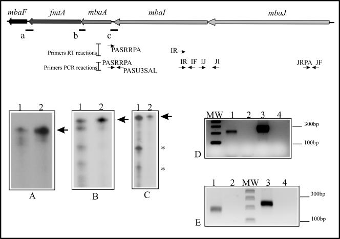
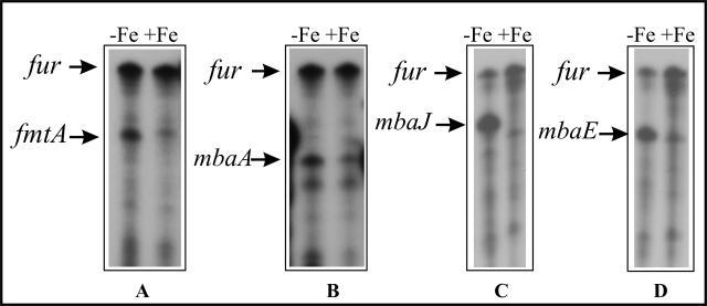
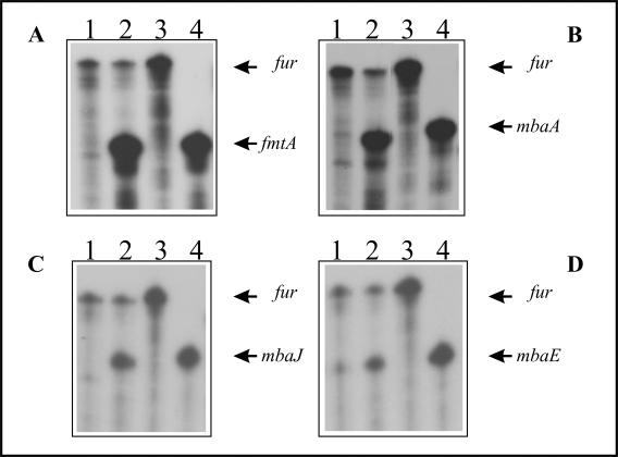
Burkholderia pseudomallei is a gram-negative facultative intracellular pathogen that causes melioidosis, an invasive disease of humans and animals. To address the response of this bacterium to iron-limiting conditions, we first performed a global transcriptional analysis of RNA extracted from bacteria grown under iron-limiting and iron-rich conditions by microarrays. We focused our study on those open reading frames (ORFs) induced under iron limitation, which encoded predicted proteins that could be involved in the biosynthesis and uptake of the siderophore malleobactin. We purified this siderophore and determined that it consisted of at least three compounds with different molecular weights. We demonstrated that ORFs BPSL1776 and BPSL1774, designated mbaA and mbaF, respectively, are involved in the biosynthesis of malleobactin, while BPSL1775, named fmtA, is involved in its transport. These genes are in an operon with two other ORFs (mbaJ and mbaI) whose transcription is under the control of MbaS, a protein that belongs to the extracytoplasmic function sigma factors. Interestingly, the transcription of the mbaA, fmtA, and mbaS genes is not controlled by the availability of the siderophore malleobactin.


Figures

FIG. 1.
FIG. 1.
Differentially expressed genes under iron-limiting conditions. (A) Genes categorized by functional classification according to TIGRFAM designations (41). Columns: 1, regulatory functions; 2, transcriptional factors; 3, DNA metabolism; 4, mobile and extrachromosomal element functions; 5, cell envelope; 6, energy metabolism; 7, cellular processes; 8, transport and binding proteins; 9, protein fate; 10, protein synthesis; 11, fatty acid and phospholipids metabolism; 12, central intermediary metabolism; 13, biosynthesis of cofactors, prosthetic groups, and carriers; 14, amino acid biosynthesis; 15, unclassified; 16, hypothetical proteins; 17, hypothetical conserved proteins. Gene regulation under iron-limiting conditions is shown as up-regulated (white columns) or down-regulated (black columns). (B) Schematic representation of the gene cluster involved in malleobactin biosynthesis and transport in B. pseudomallei K96243 and levels of induction under iron-limiting conditions. The ORF number corresponds to the genome annotation, and the proposed name is assigned to those studied in this work. Protein domains are superimposed as boxes on the mbaJ and mbaI genes that encode predicted NRPSs as follows: activation (A), condensation (C), thiolation (T), and epimerization (E). Induction levels indicate the comparison between the robust multiarray average values obtained under different growth conditions (low iron or high iron) for each transcript, following the analysis described in Materials and Methods. NC, no statistical changes observed for either growth condition.
FIG. 2.
FIG. 2.
Negative-mode electrospray ionization mass spectra of the purified siderophore malleobactin from B. pseudomallei K96243, identifying three different [M H] ion species. The fractions containing siderophore activity as assessed by bioassays were fraction I (top) and fraction II (bottom). The nominal masses (m/z) of the different ion species are indicated in the spectra.
FIG. 3.
FIG. 3.
Detection of siderophore production by B. pseudomallei on CAS agar plates. The strains were grown overnight in BHI medium, and 5 μl of the various cultures was spotted onto CAS plates with 1 mM IPTG. The plates were incubated at 37°C for 72 h. The strains are identified as follows: wt, B. pseudomallei K96243 with pMMB208; fmtA, malleobactin receptor mutant with pMMB208; fmtA/fmtA+, malleobactin receptor mutant complemented with the wild-type gene; mbaA, malleobactin biosynthesis mutant with pMMB208; mbaA/mbaA+, malleobactin biosynthesis mutant complemented with the wild-type gene; mbaS, ECF sigma factor mutant with pMMB208; mbaS/mbaS+, ECF sigma factor mutant complemented with the wild-type gene.
FIG. 4.
FIG. 4.
The mbaJIA, fmtA, and mbaF genes are transcribed together as an operon. (A to C) RNase protection assays performed with total RNA extracted from B. pseudomallei K96243 grown in Chelex-treated M9. Lanes 1, RNA from the wild type strain; lanes 2, specific probe without RNase treatment. Specific transcripts for the regions spanning the 3′ end of fmtA and the 5′ end of mbaF (primers O42 and OFRRPA), the 3′ end of mbaA and the 5′ end of fmtA (primers AORPA and OARPA), and the 5′ end of mbaA and the 3′ end of mbaI (primers PASRRPA and PASU3SAL) were detected with riboprobes a, b, and c, respectively. The solid lines below the scheme represent the locations of these riboprobes, the arrow in each panel indicates the full-length probe, and the asterisks (C) indicate the protected fragments (see the discussion in the text). (D and E) Products of RT-PCRs performed as described in Materials and Methods with primers PASRRPA (D) and IR (E). The positions of these primers are depicted below the scheme of the operon. The generated cDNAs were used as templates in PCRs with the indicated primers. (D) Lane 1, amplification with primers IF and IR; lane 3, amplification with primers PASRRPA and PASU3SAL; lanes 2 and 4, same as lanes 1 and 3 but without the addition of RT enzyme in the reaction mixtures. (E) Lane 1, amplification with primers IJ and JI; lane 3, amplification with primers JRPA and JF; lanes 2 and 4, same as lanes 1 and 3 but without the addition of RT enzyme in the reactionmixtures. MW, DNA molecular weight marker.
FIG. 5.
FIG. 5.
Expression of the mbaA, fmtA, mbaJ, and mbaE genes is regulated by the iron concentration of the medium. RNase protection assays of the mbaA, fmtA, mbaJ, and mbaE genes were performed with total RNA extracted from B. pseudomallei K96243 grown in Chelex-treated M9 (−Fe) and Chelex-treated M9 plus FAC (+Fe). (A) fmtA gene; (B) mbaA gene; (C) mbaJ gene; (D) mbaE gene. The positions of the protected RNAs are indicated by arrows. The fur gene was used as a control for RNA loading.
FIG. 6.
FIG. 6.
Expression of the mbaS gene is regulated by the iron concentration of the medium. RNase protection assays were performed with total RNA extracted from the B. pseudomallei K96243 wild-type strain and the mbaA mutant strain, grown in Chelex-treated M9 (−Fe) and Chelex-treated M9 plus FAC (+Fe) (right) and the B. pseudomallei K96243 wild-type strain and fmtA mutant strain, grown in M9 (−Fe) and M9 plus FAC (+Fe) (left). The arrows indicate the positions of the protected RNAs. The fur gene was used as a control for RNA loading.
FIG. 7.
FIG. 7.
MbaS controls the transcription of the the mbaA, fmtA, mbaJ, and mbaE genes. RNase protection assays were performed with total RNA extracted from the mbaS mutant grown in Chelex-treated M9 (lanes 1) and from the mbaS mutant complemented with the wild-type mbaS gene grown in Chelex-treated M9 plus 1 mM IPTG (lanes 2). (A) fmtA gene; (B) mbaA gene; (C) mbaJ gene; (D) mbaE gene. Lanes 3, riboprobe for the fur gene without RNase treatment; lanes 4, riboprobe for the fmtA (A), mbaA (B), mbaJ (C), and mbaE (D) genes without RNase treatment. The arrows indicate the positions of the protected RNAs. The fur gene was used as a control for RNA loading.
FIG. 8.
FIG. 8.
Transcription of the mbaA, fmtA, and mbaS genes is not controlled by malleobactin. RNase protection assays were performed with total RNA extracted from the mbaF mutant strain grown in Chelex-treated M9 plus FAC (lanes 1), Chelex-treated M9 (lanes 2), and Chelex-treated M9 plus malleobactin (lanes 3). The arrows indicate the positions of the protected RNAs. The fur gene was used as a control for RNA loading.

Similar articles

Cited by

References

    1. Altschul, S. F., T. L. Madden, A. A. Schaffer, J. Zhang, Z. Zhang, W. Miller, and D. J. Lipman. 1997. Gapped BLAST and PSI-BLAST: a new generation of protein database search programs. Nucleic Acids Res. 25:3389-3402. - PMC - PubMed
    1. Ankenbauer, R. G., and H. N. Quan. 1994. FptA, the Fe(III)-pyochelin receptor of Pseudomonas aeruginosa: a phenolate siderophore receptor homologous to hydroxamate siderophore receptors. J. Bacteriol. 176:307-319. - PMC - PubMed
    1. Atkin, C. L., J. B. Neilands, and H. J. Phaff. 1970. Rhodotorulic acid from species of Leucosporidium, Rhodosporidium, Rhodotorula, Sporidiobolus, and Sporobolomyces, and a new alanine-containing ferrichrome from Cryptococcus melibiosum. J. Bacteriol. 103:722-733. - PMC - PubMed
    1. Beare, P. A., R. J. For, L. W. Martin, and I. L. Lamont. 2003. Siderophore-mediated cell signalling in Pseudomonas aeruginosa: divergent pathways regulate virulence factor production and siderophore receptor synthesis. Mol. Microbiol. 47:195-207. - PubMed
    1. Bender, C. L., F. Alarcon-Chaidez, and D. C. Gross. 1999. Pseudomonas syringae phytotoxins: mode of action, regulation, and biosynthesis by peptide and polyketide synthetases. Microbiol. Mol. Biol Rev. 63:266-292. - PMC - PubMed

Publication types

LinkOut - more resources