. 2006 Feb 1;20(3):355-67.
doi: 10.1101/gad.1389006.

Binding of a novel SMG-1-Upf1-eRF1-eRF3 complex (SURF) to the exon junction complex triggers Upf1 phosphorylation and nonsense-mediated mRNA decay

Affiliations

Binding of a novel SMG-1-Upf1-eRF1-eRF3 complex (SURF) to the exon junction complex triggers Upf1 phosphorylation and nonsense-mediated mRNA decay

Isao Kashima et al. Genes Dev. .

Abstract

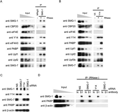
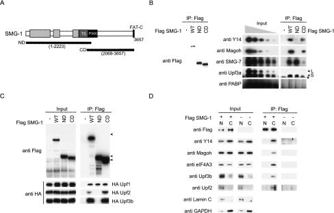
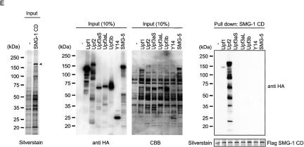
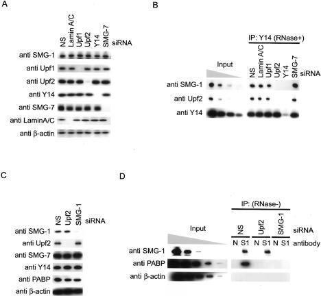
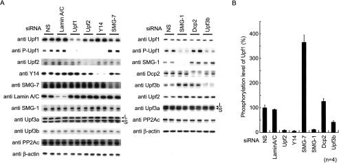
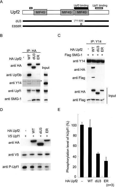
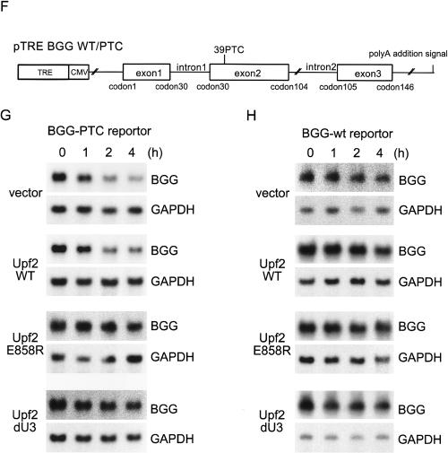
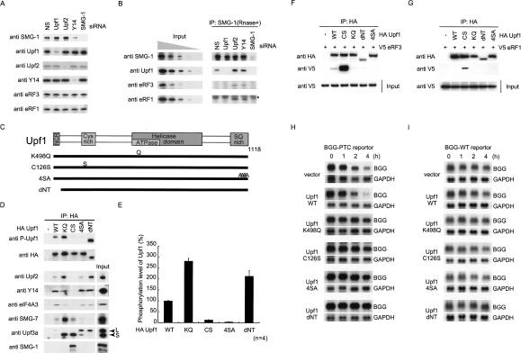
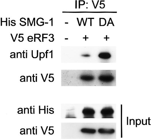
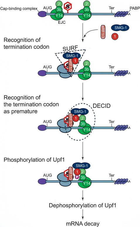
Nonsense-mediated mRNA decay (NMD) is a surveillance mechanism that degrades mRNA containing premature termination codons (PTCs). In mammalian cells, recognition of PTCs requires translation and depends on the presence on the mRNA with the splicing-dependent exon junction complex (EJC). While it is known that a key event in the triggering of NMD is phosphorylation of the trans-acting factor, Upf1, by SMG-1, the relationship between Upf1 phosphorylation and PTC recognition remains undetermined. Here we show that SMG-1 binds to the mRNA-associated components of the EJC, Upf2, Upf3b, eIF4A3, Magoh, and Y14. Further, we describe a novel complex that contains the NMD factors SMG-1 and Upf1, and the translation termination release factors eRF1 and eRF3 (SURF). Importantly, an association between SURF and the EJC is required for SMG-1-mediated Upf1 phosphorylation and NMD. Thus, the SMG-1-mediated phosphorylation of Upf1 occurs on the association of SURF with EJC, which provides the link between the EJC and recognition of PTCs and triggers NMD.


Figures

Figure 1.
Figure 1.
SMG-1 associates with the post-splicing mRNP through Y14. (A,B,D) HeLa TetOff cell lysates were immunoprecipitated with antibodies shown at the top of the figure. The immunoprecipitates (IPs), and fractionations of the cell lysates (input: 10%, 3.3%, 1.1%, and 0. 33% in A,B, and 0.11% in D of the amount immunoprecipitated) were analyzed by Western blotting with the indicated antibody probes. (C) Western blotting of siRNA-treated HeLa TetOff cell lysates. (D) N indicates the normal rabbit IgG, and S1 indicates anti-SMG-1 antibodies.
Figure 2.
Figure 2.
C-terminal half of SMG-1 directly binds to Upf2. (A) Schematic representation of SMG-1 mutants. In SMG-1-ND, the C-terminal 1589 amino acids are deleted. In SMG-1-CD, the N-terminal 2223 amino acids are deleted. The putative PIKK (PI3K-related protein kinase) and FAT-C domains are shown as black boxes. The TS (TOR-SMG-1) domains are shown as a dark-gray box. OCR (SMG-1 [one]-specific conserved region) domains are shown in light gray. (B,C) HeLa TetOff cells transfected with the plasmids shown above. A minus sign (-) indicates an empty vector; the blots were lysed and immunoprecipitated with the antibodies shown on the right (IP). IP or cell lysates (input; 10% in C, and 10%, 3.3%, 1.1%, 0. 33%, and 0.01% of the empty vector transfected in B of the amount immunoprecipitated) were then probed with the antibodies shown on the left. Arrowheads indicate the positions of SMG-1-WT, SMG-1-ND, and SMG-1-CD. SMG-1 mutants do not affect the expressions of probed proteins (data not shown). An asterisk indicates the IgG signals. (D) Immunoprecipitations and Western blotting were carried out on the nucleoplasm (N) and cytoplasm (C) of HeLa TetOff cells. Input represents 10% of the amount immunoprecipitated. (E) The SMG-1-CD region binds Upf2 in vitro. Details are described in Materials and Methods. A minus sign (-) indicates no template mRNA in an in vitro translation reaction. (Left panel) Input represents 5% of the in vitro translated reaction used for binding. Arrowheads indicate the positions of SMG-1-CD.
Figure 2.
Figure 2.
C-terminal half of SMG-1 directly binds to Upf2. (A) Schematic representation of SMG-1 mutants. In SMG-1-ND, the C-terminal 1589 amino acids are deleted. In SMG-1-CD, the N-terminal 2223 amino acids are deleted. The putative PIKK (PI3K-related protein kinase) and FAT-C domains are shown as black boxes. The TS (TOR-SMG-1) domains are shown as a dark-gray box. OCR (SMG-1 [one]-specific conserved region) domains are shown in light gray. (B,C) HeLa TetOff cells transfected with the plasmids shown above. A minus sign (-) indicates an empty vector; the blots were lysed and immunoprecipitated with the antibodies shown on the right (IP). IP or cell lysates (input; 10% in C, and 10%, 3.3%, 1.1%, 0. 33%, and 0.01% of the empty vector transfected in B of the amount immunoprecipitated) were then probed with the antibodies shown on the left. Arrowheads indicate the positions of SMG-1-WT, SMG-1-ND, and SMG-1-CD. SMG-1 mutants do not affect the expressions of probed proteins (data not shown). An asterisk indicates the IgG signals. (D) Immunoprecipitations and Western blotting were carried out on the nucleoplasm (N) and cytoplasm (C) of HeLa TetOff cells. Input represents 10% of the amount immunoprecipitated. (E) The SMG-1-CD region binds Upf2 in vitro. Details are described in Materials and Methods. A minus sign (-) indicates no template mRNA in an in vitro translation reaction. (Left panel) Input represents 5% of the in vitro translated reaction used for binding. Arrowheads indicate the positions of SMG-1-CD.
Figure 3.
Figure 3.
Association between SMG-1 and mRNP requires Upf2. (A,C) HeLa TetOff cell lysates transfected with the siRNAs shown above the blots were analyzed by Western blotting with the indicated antibodies as probes. (B,D) HeLa TetOff cell lysates transfected with siRNAs were immunoprecipitated with anti-Y14 antibody in the presence of RNase (B), or with anti-SMG-1 or normal rabbit IgG in the absence of RNase (D; IP). IP or cell lysates (input; 10%, 3.3%, 1.1%, and 0. 33% in B, and 0.11% in D) were then probed with the antibodies shown on the left.
Figure 4.
Figure 4.
SMG-1-mediated phosphorylation of Upf1 requires Upf2, Upf3b, and Y14 in vivo. (A) HeLa TetOff cell lysates transfected with the siRNAs shown above the blots were analyzed by Western blotting with the indicated antibody probes. Anti-P-Upf1 antibody recognizes phosphorylated S1078 and S1096 residues. Arrowheads indicate the positions of Upf3aL and Upf3aS. (B) Quantification of the phosphorylation level of Upf1 in vivo when NMD components were depleted.
Figure 5.
Figure 5.
Interaction between Upf2 and Upf3b is required for the association between SMG-1 and mRNP during NMD. (A) Schematic representation of Upf2 mutants. The solid line indicates the Upf3-binding region. The white line indicates the Upf1-binding region. MIF4G (mammalian eIF4G like) domains are shown as dark-gray boxes. (B) Upf2 mutants do not interact with Upf3b and Y14. Ectopic HA-Upf2-WT, HA-Upf2-dU3, or HA-Upf2-E858R expressed in HeLa TetOff cells was immunoprecipitated under the conditions of RNase-treatment and analyzed for the presence of endogenous proteins by Western blotting with the indicated antibody probes. A minus sign (-) indicates an empty vector. Upf2 mutants do not affect expressions of probed proteins (data not shown). Input represents 10% of the amount immunoprecipitated and of the empty vector transfected. An asterisk indicates the IgG signals. (C) HeLa TetOff cell lysates transfected with plasmids were immunoprecipitated with anti-Y14 antibody under conditions of RNase-treatment (IP). IP and the input fraction (10% of the amount immunoprecipitated) were analyzed by Western blotting with the indicated antibody probes. (D) Overexpression of Upf2 mutants inhibits the phosphorylation of Upf1. (E) Quantification of the phosphorylation level of Upf1 when Upf2 mutants were overexpressed. (F) Schematic representation of human β-globin gene (BGG) reporter constructs BGG-WT and BGG-PTC. The ORF is represented by boxes and introns and UTRs are represented by lines. (G,H) Overexpression of Upf2-dU3 and Upf2-E852R inhibit PTC-containing β-globin mRNA decay. HeLa TetOff cells were transfected with a reporter plasmid (BGG-WT or BGG-PTC). Cells were simultaneously transfected with the Upf2 plasmids, as indicated on the left of blots. Panels show typical examples of the results of Northern blotting. The quantities of BGG mRNA, normalized to GADPH signals, are graphed in Supplementary Figure 5.
Figure 5.
Figure 5.
Interaction between Upf2 and Upf3b is required for the association between SMG-1 and mRNP during NMD. (A) Schematic representation of Upf2 mutants. The solid line indicates the Upf3-binding region. The white line indicates the Upf1-binding region. MIF4G (mammalian eIF4G like) domains are shown as dark-gray boxes. (B) Upf2 mutants do not interact with Upf3b and Y14. Ectopic HA-Upf2-WT, HA-Upf2-dU3, or HA-Upf2-E858R expressed in HeLa TetOff cells was immunoprecipitated under the conditions of RNase-treatment and analyzed for the presence of endogenous proteins by Western blotting with the indicated antibody probes. A minus sign (-) indicates an empty vector. Upf2 mutants do not affect expressions of probed proteins (data not shown). Input represents 10% of the amount immunoprecipitated and of the empty vector transfected. An asterisk indicates the IgG signals. (C) HeLa TetOff cell lysates transfected with plasmids were immunoprecipitated with anti-Y14 antibody under conditions of RNase-treatment (IP). IP and the input fraction (10% of the amount immunoprecipitated) were analyzed by Western blotting with the indicated antibody probes. (D) Overexpression of Upf2 mutants inhibits the phosphorylation of Upf1. (E) Quantification of the phosphorylation level of Upf1 when Upf2 mutants were overexpressed. (F) Schematic representation of human β-globin gene (BGG) reporter constructs BGG-WT and BGG-PTC. The ORF is represented by boxes and introns and UTRs are represented by lines. (G,H) Overexpression of Upf2-dU3 and Upf2-E852R inhibit PTC-containing β-globin mRNA decay. HeLa TetOff cells were transfected with a reporter plasmid (BGG-WT or BGG-PTC). Cells were simultaneously transfected with the Upf2 plasmids, as indicated on the left of blots. Panels show typical examples of the results of Northern blotting. The quantities of BGG mRNA, normalized to GADPH signals, are graphed in Supplementary Figure 5.
Figure 6.
Figure 6.
The SMG-1-Upf1 complex with eRF1 and eRF3. (A) HeLa TetOff cell lysates transfected with the siRNAs shown above the blots were analyzed by Western blotting with the indicated antibody probes. (B) HeLa TetOff cell lysates transfected with siRNA were immunoprecipitated with anti-SMG-1 antibodies (IP). IP or cell lysates (input; 10%, 3.3%, 1.1%, 0. 33%, and 0.11% of the amount immunoprecipated) were then probed with the antibodies shown on the left. An asterisk indicates the IgG signals. (C) Schematic structures of human Upf1 mutants. The metazoan-specific N-terminal conserved region (NCR), cysteine-rich regions, helicase domains, and SQ-rich region of Upf1 are shown in gray boxes. (D,F,G) HeLa TetOff cells transfected with the plasmids indicated above each blot; a minus sign (-) indicates an empty vector. Cell lysates (input) were immunoprecipitated with the antibodies under the conditions of RNase-treatment shown on the top and probed with the antibodies shown on the left. Upf1 mutants do not affect expressions of probed proteins (data not shown). Input represents 10% of the amount immunoprecipitated. (D) Immunoprecipitations of Upf1 mutants. (E) Quantification of the phosphorylation level of Upf1 mutants. (F,G) Upf1-C126S strongly interact with eRF1 and eRF3. (H,I) Similar to Figure 5G, HeLa TetOff cells were transfected with a reporter plasmid (BGG-WT or BGG-PTC). Cells were simultaneously transfected with the Upf1 plasmids, as indicated on the left. Panels show typical examples of the results of Northern blotting. The quantities of BGG mRNA, normalized to GADPH signals, are graphed in Supplementary Figure 5.
Figure 7.
Figure 7.
The interaction between Upf1 and eRF3 increased upon overexpression of His-SMG-1-DA. HeLa TetOff cell lysates transfected with the plasmids shown above. Cell lysates (input) were immunoprecipitated with the antibodies under the conditions of RNase-treatment as shown on the top and were probed with the antibodies shown on the left. Input represents 10% of the amount immunoprecipitated.
Figure 8.
Figure 8.
A model illustrating the SMG-1-mediated phosphorylation of Upf1 that occurs on post-splicing mRNP after translation termination. Details are described in the text. (P) Phosphate group; (1) Upf1; (2) Upf2; (3b) Upf3b; (eRF1/3) eRF1-eRF3 complex; (EJC) exon junction complex; (AUG) start codon; (Ter) termination codon; (S) stop codon; (SURF) SMG-1-Upf1-eRF1-eRF3 complex; (DECID) decay-inducing complex.

Comment in

Similar articles

Cited by

References

    1. Anders K.R., Grimson, A., and Anderson, P. 2003. SMG-5, required for C. elegans nonsense-mediated mRNA decay, associates with SMG-2 and protein phosphatase 2A. EMBO J. 22: 641-650. - PMC - PubMed
    1. Baker K.E. and Parker, R. 2004. Nonsense-mediated mRNA decay: Terminating erroneous gene expression. Curr. Opin. Cell Biol. 16: 293-299. - PubMed
    1. Cali B.M. and Anderson, P. 1998. mRNA surveillance mitigates genetic dominance in Caenorhabditis elegans. Mol. Gen. Genet. 260: 176-184. - PubMed
    1. Cali B.M., Kuchma, S.L., Latham, J., and Anderson, P. 1999. smg-7 is required for mRNA surveillance in Caenorhabditis elegans. Genetics 151: 605-616. - PMC - PubMed
    1. Chan C.C., Dostie, J., Diem, M.D., Feng, W., Mann, M., Rappsilber, J., and Dreyfuss, G. 2004. eIF4A3 is a novel component of the exon junction complex. RNA 10: 200-209. - PMC - PubMed

Publication types

MeSH terms