. 2006 Feb 1;20(3):368-79.
doi: 10.1101/gad.371406.

Sex-lethal imparts a sex-specific function to UNR by recruiting it to the msl-2 mRNA 3' UTR: translational repression for dosage compensation

Affiliations

Sex-lethal imparts a sex-specific function to UNR by recruiting it to the msl-2 mRNA 3' UTR: translational repression for dosage compensation

Kent Duncan et al. Genes Dev. .

Abstract

MSL-2 (male-specific lethal 2) is the limiting component of the Drosophila dosage compensation complex (DCC) that specifically increases transcription from the male X chromosome. Ectopic expression of MSL-2 protein in females causes DCC assembly on both X chromosomes and lethality. Inhibition of MSL-2 synthesis requires the female-specific protein sex-lethal (SXL), which binds to the msl-2 mRNA 5' and 3' untranslated regions (UTRs) and blocks translation through distinct UTR-specific mechanisms. Here, we purify translationally silenced msl-2 mRNPs and identify UNR (upstream of N-ras) as a protein recruited to the 3' UTR by SXL. We demonstrate that SXL requires UNR as a corepressor for 3'-UTR-mediated regulation, imparting a female-specific function to the ubiquitously expressed UNR protein. Our results reveal a novel functional role for UNR as a translational repressor and indicate that UNR is a key component of a "fail-safe" dosage compensation regulatory system that prevents toxic MSL-2 synthesis in female cells.


Figures
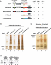
Figure 1.
Figure 1.
GRNA chromatography identifies CG7015/Drosophila UNR as a candidate msl-2 mRNA translational corepressor. (A) RNAs used for GRNA chromatography are depicted schematically and their expected associations are indicated. (B) RNAs were incubated in Drosophila embryo translation extracts to assemble silenced RNPs (lanes 2,4) or control nonsilenced RNPs (lanes 3,5) and were subsequently purified by GRNA chromatography. Lane 1 contains the negative control BSEF RNA without the box-B sequence. Eluates were separated by SDS-PAGE and silver stained to identify bands unique to silenced RNPs. Each silenced RNP and corresponding nonsilenced control lane was divided into ∼20 subregions, and peptides from each were identified by mass spectrometry. (C) Sucrose density gradient fractionation of RNPs was performed and fractions containing repressed heavy mRNPs (Beckmann et al. 2005) (lane 2) or corresponding fractions from control samples (lanes 1,3) were pooled prior to GRNA chromatography. Arrowheads indicate CG7015/UNR, λ-GST, and SXL. (D) BSEF-box-B (lanes 3,4) or BmS(EF)m-box-B (lanes 5,6) RNAs were incubated in embryo extracts in the presence or absence of added recombinant SXL as indicated. GRNA eluates were normalized for RNA isolation efficiency and analyzed by SDS-PAGE and immunoblotting with anti-UNR antibodies.
Figure 2.
Figure 2.
ORF CG7015 is Drosophila UNR. ORF CG7015 was aligned with UNR orthologs from a variety of species using ClustalW (EBI). Darker color indicates a higher degree of conservation of a specific residue. The five CSDs are underlined in green (CSD-1-CSD-5). Red boxes highlight the FFH sequence in the RNP-2 consensus that is a signature of UNR CSDs.
Figure 3.
Figure 3.
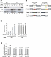
Endogenous SXL in Kc cells represses msl-2 mRNA translation via 5′- and 3′-UTR SXL-binding sites. (A) Relative levels of MSL-2, SXL, or control α-tubulin in whole-cell lysates from SL-2 cells (lanes 1-3), Kc cells (lanes 4-6), control GFP dsRNA-treated Kc cells (lane 7), or SXL dsRNA-treated cells (lane 8) were analyzed by immunoblotting. (B) msl-2-β-gal reporter constructs are depicted schematically. The HSP70 promoter and SV40 poly(A) sequence that combine to give low-level constitutive expression are indicated. Solid black lines represent sequences derived from the msl-2 5′ and 3′ UTRs, as indicated. SXL-binding sites A-F are shown as green ovals. Region 2456 is shown in red. (C) Relative β-gal activity of Kc cells treated with the indicated dsRNAs and transfected with the indicated reporter constructs. All values were normalized to Renilla luciferase (Rluc) activity from a cotransfected plasmid to correct for any variations in transfection efficiency. Wild-type (WT) construct average activity in control GFP RNAi cells was set to 1.0. Average values and standard deviations in at least four independent RNAi experiments are shown. (D) Relative mRNA levels for transfected samples were analyzed by qPCR. msl-2-β-gal reporter values were normalized to Rluc mRNA levels. For each reporter, levels in SXL-depleted samples are shown relative to levels in the corresponding control GFP dsRNA-treated cells. Average values and standard deviations correspond to at least three independent RNAi experiments used for reporter activity analysis in C.
Figure 4.
Figure 4.
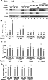
UNR acts as a 3′-UTR corepressor in conjunction with SXL. (A) Whole-cell extracts were prepared from mock-treated SL-2 cells (lanes 1-3), mock-treated Kc cells (lanes 4-6), or Kc cells treated with dsRNAs targeting EGFP (lane 7), SXL (lane 8), UNR (lane 9) alone, or in combination with SXL dsRNAs (lanes 10-12, respectively). Relative levels of UNR, SXL, and control α-tubulin were determined by immunoblotting. (B,C) Kc cells were treated with the indicated dsRNAs and subsequently transfected with wild-type (WT), 5′ mut, 3′ mut, or 5′ + 3′ mut reporters, as indicated. Relative β-gal activities were normalized to Rluc as in Figure 3C. For each reporter, the level of expression in GFP-treated cells is set to 1.0, and the fold increase in expression in the UNR and/or SXL RNAi-treated cells is shown. Average values and standard deviations in at least four (B) or two (C) independent RNAi experiments are shown. (D) Relative mRNA levels in the transfected samples were analyzed by qPCR as in Figure 3. For each reporter, the mRNA level in UNR-treated samples is shown relative to the amount in the corresponding GFP-treated samples, which is set to 1.0. Average values and standard deviations in at least three independent RNAi experiments are shown.
Figure 5.
Figure 5.
SXL recruits UNR for a sex-specific function. (A) Relative levels of msl-2-β-gal reporter activity in SL-2 cells treated with the indicated dsRNAs and transfected with either wild type (WT, light bars) or 5′ + 3′ mut (dark bars) reporters in the absence of SXL. Rluc-normalized activities of both reporters are compared with the 5′ + 3′ mut reporter, which is set to 100% for each dsRNA treatment. (B) Relative reporter activities in cells cotransfected with pHSP70-SXL. (C) UNR and α-tubulin levels in duplicate samples from wild-type (WT) reporter-transfected, UNR dsRNA-treated (lanes 1-4), or EGFP dsRNA-treated (lanes 5-8) cells were analyzed by immunoblotting. (D) RNase Protection Assay analysis of relative mRNA levels in untreated cells (lanes 1-9) or UNR dsRNA-treated cells (lanes 10-13), transfected with an actin5C promoter-LacZ-positive control (lanes 1,2), no plasmid (lanes 3-5), wild-type (WT) reporter (lanes 6,8,10,12), or 5′ +3′ mut reporter (lanes 7,9,11,13).
Figure 6.
Figure 6.
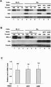
UNR is required for translational repression of endogenous msl-2 mRNA in Kc cells. (A) Levels of UNR, SXL, and α-tubulin protein in whole-cell lysates were measured by immunoblotting. Mock RNAi SL-2 cells (lanes 1-3), mock RNAi Kc cells (lanes 4-6), or Kc cells treated with dsRNAs targeting EGFP (lane 7), SXL (lane 8), or UNR (lane 9). (B) Extracts from A were analyzed for MSL-2, SXL, and α-tubulin protein levels by immunoblotting. (Lanes 1-3) Untreated SL-2 cells. (Lanes 4-6) SXL dsRNA-treated Kc cells. (Lane 7) Untreated Kc cells. (Lane 8) Kc cells treated with dsRNAs targeting GFP. (Lane 9) Kc cells treated with dsRNAs targeting UNR. Blots shown are representative of multiple blots analyzing samples from three independent RNAi experiments. (C) Levels of endogenous msl-2 mRNA in samples from Kc cells treated with the indicated dsRNAs were analyzed by qPCR. Average values and standard deviations in four independent RNAi experiments are shown.
Figure 7.
Figure 7.
SXL imparts a novel sex- and mRNA-specific translational repressor function to UNR. A model for UNR's sex-specific role in translational regulation of MSL-2 synthesis for dosage compensation is shown. In female cells, SXL first recruits UNR to the 3′ UTR of msl-2 mRNA. Subsequently, 3′-UTR-bound UNR, possibly in concert with SXL and/or other factors, acts as a translational corepressor, inhibiting recruitment of the small ribosomal subunit-containing 43S preinitiation complex to the mRNA 5′ end. 43S complexes that escape this primary translational block are challenged by the UNR-independent scanning block mediated by 5′-UTR-bound SXL (Beckmann et al. 2005). The dual inhibitory mechanisms combine to prevent toxic expression of MSL-2 in females. In male cells, where MSL-2 translation is essential for dosage compensation, UNR and msl-2 mRNA are both present at high levels. The absence of SXL prevents stable interaction of UNR with the msl-2 mRNA, allowing active msl-2 mRNA translation.

Similar articles

Cited by

References

    1. Abaza I., Coll, O., Patalano, S., and Gebauer, F. 2006. Drosophila UNR is required for translational repression of male-specific-lethal 2 mRNA during regulation of X-chromosome dosage compensation. Genes & Dev. (this issue). - PMC - PubMed
    1. Arbeitman M.N., Furlong, E.E., Imam, F., Johnson, E., Null, B.H., Baker, B.S., Krasnow, M.A., Scott, M.P., Davis, R.W., and White, K.P. 2002. Gene expression during the life cycle of Drosophila melanogaster. Science 297: 2270-2275. - PubMed
    1. Ausubel F.M., Brent, R., Kingston, R.E., Moore, D.D., Seidman, J.G., Smith, J.A., and Struhl, K. 1999. Short protocols in molecular biology. John Wiley & Sons, Inc., New York.
    1. Baker B.S., Gorman, M., and Marin, I. 1994. Dosage compensation in Drosophila. Annu. Rev. Genet. 28: 491-521. - PubMed
    1. Bashaw G.J. and Baker, B.S. 1995. The msl-2 dosage compensation gene of Drosophila encodes a putative DNA-binding protein whose expression is sex specifically regulated by Sex-lethal. Development 121: 3245-3258. - PubMed

Publication types

MeSH terms