. 2006 Feb 1;20(3):380-9.
doi: 10.1101/gad.371906.

Drosophila UNR is required for translational repression of male-specific lethal 2 mRNA during regulation of X-chromosome dosage compensation

Affiliations

Drosophila UNR is required for translational repression of male-specific lethal 2 mRNA during regulation of X-chromosome dosage compensation

Irina Abaza et al. Genes Dev. .

Abstract

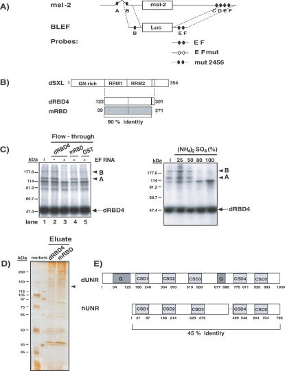
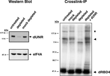
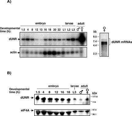
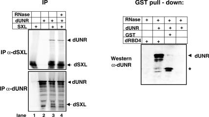
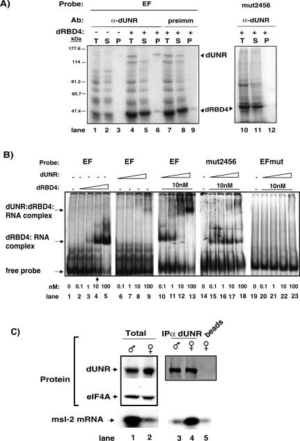
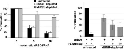
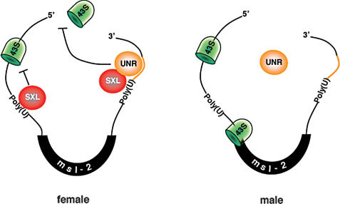
The inhibition of male-specific lethal 2 (msl-2) mRNA translation by the RNA-binding protein sex-lethal (SXL) is an essential regulatory step for X-chromosome dosage compensation in Drosophila melanogaster. The mammalian upstream of N-ras (UNR) protein has been implicated in the regulation of mRNA stability and internal ribosome entry site (IRES)-dependent mRNA translation. Here we have identified the Drosophila homolog of mammalian UNR as a cofactor required for SXL-mediated repression of msl-2 translation. UNR interacts with SXL, a female-specific protein. Although UNR is present in both male and female flies, binding of SXL to uridine-rich sequences in the 3' untranslated region (UTR) of msl-2 mRNA recruits UNR to adjacent regulatory sequences, thereby conferring a sex-specific function to UNR. These data identify a novel regulator of dosage compensation in Drosophila that acts coordinately with SXL in translational control.


Figures

Figure 1.
Figure 1.
Purification of Drosophila UNR. (A) Schematic representation of the msl-2 mRNA and the RNA constructs used in this study. msl-2 mRNA contains SXL-binding sites (A-F, filled ovals) in its 5′ UTR (626 nucleotides [nt]) and 3′ UTR (1047 nt). BLEF mRNA harbors the minimal msl-2 sequences required for translational repression, consisting of 69 nt in the 5′ UTR containing site B, and 46 nt in the 3′ UTR containing sites E and F, fused to the Firefly luciferase ORF (Gebauer et al. 2003). Probes used for UV-cross-link and gel mobility-shift assays are also depicted (see Materials and Methods for their detailed sequences). (B) Scheme of the Drosophila SXL protein (dSXL) and its derivatives. dRBD4 is a deletion derivative fully functional in translational repression; mRBD is the equivalent fragment of the SXL homolog from Musca domestica, which shows no translational repression ability (Grskovic et al. 2003). Amino acid numbers are indicated, as well as the percentage of identity between the Drosophila and Musca SXL fragments. (C, left panel) Retention of putative corepressors in SXL columns. Embryo extract was loaded on glutathione-Sepharose columns containing either GST-dRBD4, GST-mRBD, or GST alone, in the presence or absence of the EF RNA fragment. Retention of the polypeptides A and B was tested by analyzing their presence in the column flowthrough using UV-cross-link to radiolabeled EF RNA and coimmunoprecipitation with dRBD4, as previously described (Grskovic et al. 2003). (i) Input. (C, right panel) Enrichment of the putative corepressors by ammonium sulfate precipitation. Drosophila embryo extracts were subjected to precipitation by saturation with (NH4)2SO4. The polypeptides A and B (arrowheads) were detected by the cross-link-IP assay described above. (D) Enrichment of an ∼130-kDa protein in the dRBD4 column eluate. Proteins present in the eluates from dRBD4 and mRBD columns were separated in a 10% acrylamide gel and silver stained (see Results for further experimental details). The putative ∼130-kDa corepressor is indicated with an arrow. (E) Schematic diagram of Drosophila and human UNR proteins (dUNR and hUNR, respectively). dUNR contains five CSDs and two glutamine-rich regions (Q). The amino acid numbers and the identity between the two UNR proteins are shown.
Figure 2.
Figure 2.
dUNR interacts with msl-2 mRNA. Antibodies against dUNR were generated and used to deplete Drosophila embryo extracts. (Left panel) The extent of dUNR depletion was assessed by Western blot using eIF4A as a specificity control. (Right panel) The presence of the polypeptides A and B in the depleted extracts was tested by cross-link-IP, as indicated in the legend of Figure 1. The assay was performed in the absence or presence of 100 ng of purified recombinant dUNR. The arrowhead indicates the position of dUNR, while the asterisk indicates a complex between dUNR and dRBD4.
Figure 3.
Figure 3.
Developmental distribution of dUNR. (A, left panel) Semiquantitative RT-PCR analysis of dUNR mRNA during development. Amplification of actin mRNA within the same reactions was performed as a control. Amplified fragments were visualized by Southern blot. (A, right panel) Northern blot of female RNA using the dUNR full-length ORF as a probe. Molecular weight markers are indicated on the left. (B) Western blot of dUNR during Drosophila development. The amount of eIF4A was monitored as a loading control.
Figure 4.
Figure 4.
Immunostaining of dUNR. Blastoderm-stage Drosophila embryos were incubated with preimmune serum (A) or α-dUNR serum (B-D) (green). DNA was stained using TOPRO3 (blue). Identical exposures were done for all pictures. The arrowhead indicates the stronger fluorescence intensity around the nucleus in somites (C) and pole cells (D).
Figure 5.
Figure 5.
dUNR interacts with dSXL. (Left panel) Coimmunoprecipitation of in vitro-translated dUNR and dSXL proteins with either α-dSXL or α-dUNR antibodies, in the absence or presence of RNase A. (Right panel) Pull-down of purified recombinant dUNR with glutathione-agarose beads containing either GST or GST-dRBD4. The assay was performed in the presence of RNase A. The position of dUNR is indicated. The asterisk denotes a bacterial contaminant present in the GST preparation that cross-reacts with the α-dUNR antibody.
Figure 6.
Figure 6.
Binding of dUNR to msl-2 mRNA requires dSXL. (A) Cross-link of endogenous dUNR to 32P-labeled EF RNA, and IP with α-dUNR antibodies. The total set of cross-linked proteins (T), as well as the supernatant (S) and pellet (P) fractions of the IP are shown. In some tubes, dRBD4 was added to the reaction before cross-linking. Precipitation with preimmune serum, and cross-linking with radiolabeled mut2456 RNA (lacking dUNR-binding sites) were carried as negative controls. The positions of dRBD4 and dUNR are indicated. (B) Gel mobility-shift assays of radiolabeled wild-type EF RNA or derivatives lacking either dSXL-binding sites (EFmut) or dUNR-binding sites (mut2456), in the absence or presence of increasing amounts of recombinant dUNR and dRBD4 proteins. The positions of the different complexes are indicated. (C) dUNR interacts with msl-2 mRNA in female, but not male, flies. (Upper panel) Western blot of dUNR in total extracts from adult male and female flies (lanes 1,2), and after IP with α-dUNR serum (lanes 3,4) or beads alone (lane 5). The amount of eIF-4A was measured as a loading control. (Lower panel) Semiquantitative RT-PCR analysis of msl-2 mRNA from total (1 μg) male and female RNA (lanes 1,2) and equivalent volumes of RNA extracted from the IP shown in the upper panel (lanes 3-5). Amplified fragments were visualized by Southern blot.
Figure 7.
Figure 7.
Drosophila UNR is required for translational repression of msl-2 mRNA. (Left panel) BLEF mRNA was incubated in typical translation reactions containing increasing amounts of dRBD4 and either untreated (black bars), dUNR-depleted (gray bars), or mock-depleted (white bars) extracts. Firefly luciferase values were corrected for cotranslated Renilla expression and plotted against the molar ratio dRBD4/RNA. The activity obtained in the absence of recombinant protein was taken as 100%. (Right panel) Translation inhibition by dSXL is restored upon addition of recombinant dUNR. Translation reactions were assembled with dUNR-depleted extracts in the absence or presence of increasing amounts of recombinant dUNR. Untreated extracts were used as a reference (black bars). Where indicated, 20-fold molar excess of dRBD4 over reporter BLEF RNA was used.
Figure 8.
Figure 8.
Model for the role of Drosophila UNR in the regulation of msl-2 mRNA translation. The female-specific protein SXL binds to poly(U) stretches in the 5′ and 3′ UTRs of msl-2 mRNA. SXL bound to the 3′ UTR recruits UNR to adjacent purine-rich sequences. The SXL/UNR complex inhibits the association of the 43S ribosomal complex with the 5′ end of the mRNA, thereby repressing translation. SXL bound to the 5′ UTR inhibits the scanning of ribosomal complexes that may have escaped the UNR block (Beckmann et al. 2005). In male flies, which lack SXL, UNR does not bind to the 3′ UTR of msl-2 mRNA and translation proceeds.

Similar articles

Cited by

References

    1. Arbeitman M.N., Furlong, E.E., Imam, F., Johnson, E., Null, B.H., Baker, B.S., Krasnow, M.A., Scott, M.P., Davis, R.W., and White, K.P. 2002. Science 297: 2270-2275. - PubMed
    1. Bashaw G.J. and Baker, B.S. 1997. The regulation of the Drosophila msl-2 gene reveals a function for Sex-lethal in translational control. Cell 89: 789-798. - PubMed
    1. Beckmann K., Grskovic, M., Gebauer, F., and Hentze, M.W. 2005. A dual inhibitory mechanism for ribosomal 43S complex recruitment and scanning restricts msl-2 mRNA translation for dosage compensation in Drosophila. Cell 122: 529-540. - PubMed
    1. Belote J.M. 1983. Male-specific lethal mutations of Drosophila melanogaster. II. Parameters of gene action during male development. Genetics 105: 881-896. - PMC - PubMed
    1. Bopp D., Bell, L.R., Cline, T.W., and Schedl, P. 1991. Developmental distribution of female-specific Sex-lethal proteins in Drosophila melanogaster. Genes & Dev. 5: 403-415. - PubMed

Publication types

MeSH terms