. 2006 Apr;17(4):1632-42.
doi: 10.1091/mbc.e05-10-0912. Epub 2006 Feb 1.

Loss of P4 ATPases Drs2p and Dnf3p disrupts aminophospholipid transport and asymmetry in yeast post-Golgi secretory vesicles

Affiliations

Loss of P4 ATPases Drs2p and Dnf3p disrupts aminophospholipid transport and asymmetry in yeast post-Golgi secretory vesicles

Nele Alder-Baerens et al. Mol Biol Cell. 2006 Apr.

Abstract

Eukaryotic plasma membranes generally display asymmetric lipid distributions with the aminophospholipids concentrated in the cytosolic leaflet. This arrangement is maintained by aminophospholipid translocases (APLTs) that use ATP hydrolysis to flip phosphatidylserine (PS) and phosphatidylethanolamine (PE) from the external to the cytosolic leaflet. The identity of APLTs has not been established, but prime candidates are members of the P4 subfamily of P-type ATPases. Removal of P4 ATPases Dnf1p and Dnf2p from budding yeast abolishes inward translocation of 6-[(7-nitrobenz-2-oxa-1,3-diazol-4-yl)aminocaproyl] (NBD)-labeled PS, PE, and phosphatidylcholine (PC) across the plasma membrane and causes cell surface exposure of endogenous PE. Here, we show that yeast post-Golgi secretory vesicles (SVs) contain a translocase activity that flips NBD-PS, NBD-PE, and NBD-PC to the cytosolic leaflet. This activity is independent of Dnf1p and Dnf2p but requires two other P4 ATPases, Drs2p and Dnf3p, that reside primarily in the trans-Golgi network. Moreover, SVs have an asymmetric PE arrangement that is lost upon removal of Drs2p and Dnf3p. Our results indicate that aminophospholipid asymmetry is created when membrane flows through the Golgi and that P4-ATPases are essential for this process.


Figures
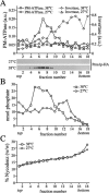
Figure 1.
Figure 1.
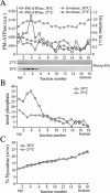
Gradient fractionation of post-Golgi SVs accumulated in 38°C-shifted sec6-4 cells. Membranes enriched in SVs were prepared from 38°C-shifted or 27°C-grown sec6-4 cells by differential centrifugation, loaded on the bottom of a linear Nycodenz/sorbitol gradient, and then floated to equilibrium by centrifugation. (A) Fractions were collected from the top and analyzed for enzyme activities and by immunoblotting. Enzyme activities were determined as described in Materials and Methods and expressed in arbitrary units (a.u.) based on the absorbance measured at 660 nm (PM-ATPase) or 540 nm (invertase). Immunoblots were probed with a mAb against the HA epitope to detect HA-tagged Pma1p. (B) Lipid phosphorus content per fraction was determined as described in Materials and Methods. (C) Fraction densities were measured by reading refractive indices on a Bausch & Lomb refractometer.
Figure 2.
Figure 2.
SVs accumulated in 38°C-shifted sec6-4 cells contain NBD-phospholipid translocase activity. (A) Membranes enriched in SVs were prepared from 38°C-shifted sec6-4 cells by differential centrifugation, labeled with NBD-lipids, and incubated for 3 h at 25°C without ATP. The reaction was then split, ATP was added to one-half, and the incubation was continued an additional 2 h. At times indicated, the fraction of fluorescent lipid remaining in the cytosolic leaflet was determined by back-extraction using defatted BSA as described in Materials and Methods. Data shown are means ± SD of three independent experiments. (B) SV-enriched membranes prepared from 27°C-grown sec6-4 cells were analyzed for ATP-dependent NBD-lipid transport by BSA back-extraction as in A. Data shown are means ± SD of two independent experiments. Note that the membranes from 27°C-grown cells display an apparent reduction in the passive movement of all four NBD-lipids compared with membranes from 38°C-shifted cells. Given the assay conditions used, this may be explained by an overall increase in the rate of flip-flop because of a larger fraction of ER membranes in the 27°C membrane preparation (also see Figure 6A).
Figure 3.
Figure 3.
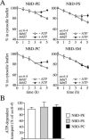
SV-associated phospholipid translocase activity is independent of Dnf1p and Dnf2p. (A) SV-enriched membranes were prepared from 38°C-shifted sec6-4Δdnf1Δdnf2 cells, labeled with NBD-lipids, and incubated for 3 h at 25°C without ATP. The reaction was then split, ATP was added to one-half, and the incubation was continued an additional 2 h. At times indicated, the fraction of fluorescent lipid remaining in the cytosolic leaflet was determined as in the legend to Figure 2. (B) As described in A, except that membranes were labeled with NBD-lipid and then incubated for 4 h in the continuous presence or absence of ATP. The level of ATP-dependent lipid transport was calculated by subtracting the accessible pool of fluorescent lipid in membranes incubated without ATP from that in ATP-incubated membranes and expressed as percentage of transport measured in membranes from 38°C-shifted sec6-4 cells. Data shown are means ± SD of at least two independent experiments.
Figure 4.
Figure 4.
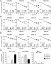
Loss of Drs2p and Dnf3p abolishes SV-associated phospholipid translocase activity. SV-enriched membranes prepared from 38°C-shifted sec6-4Δdrs2 (A), sec6-4Δdnf3 (B) and sec6-4Δdrs2Δdnf3 cells (C) were labeled with NBD-lipids and incubated for 3 h at 25°C without ATP. The reaction was then split, ATP was added to one-half, and the incubation was continued an additional 2 h. At times indicated, the fraction of fluorescent lipid remaining in the cytosolic leaflet was determined as in the legend to Figure 2. (D) SV-enriched membranes from sec6-4Δdrs2, sec6-4Δdnf3, and sec6-4Δdrs2Δdnf3 cells were labeled with NBD-lipid and then incubated for 4 h in the continuous presence or absence of ATP. The level of ATP-dependent lipid transport was calculated by subtracting the accessible pool of fluorescent lipid in membranes incubated without ATP from that in ATP-incubated membranes and expressed as percentage of transport measured in membranes from 38°C-shifted sec6-4 cells. Data shown are means ± SD of at least two independent experiments.
Figure 5.
Figure 5.
Formation of Pma1p-transporting SVs is independent of Drsp2 and Dnf3p. SV-enriched membranes were prepared from 38°C-shifted or 27°C-grown sec6-4Δdrs2Δdnf3 cells by differential centrifugation, loaded on the bottom of a linear Nycodenz/sorbitol gradient and then floated to equilibrium by centrifugation. (A) Fractions were collected from the top and analyzed for enzyme activities and by immunoblotting as in the legend to Figure 1. (B) Lipid phosphorus content was determined as described in Materials and Methods. (C) Fraction densities were measured by reading refractive indices on a Bausch & Lomb refractometer.
Figure 6.
Figure 6.
SV-associated translocase activity from a Dnf1,2,3-deficient mutant recognizes NBD-labeled PS and PE but not NBD-PC. SV-enriched membranes prepared from 38°C-shifted sec6-4Δdrs2Δdnf1,2,3 mutant cells expressing Drs2p from a multicopy (2μ) plasmid were labeled with NBD-lipids and incubated for 3 h at 25°C without ATP. The reaction was then split, ATP was added to one-half, and the incubation was continued an additional 2 h. At times indicated, the fraction of fluorescent lipid remaining in the cytosolic leaflet was determined as in the legend to Figure 2. Data shown are means ± SD of two independent experiments.
Figure 7.
Figure 7.
Pma1p-transporting SVs contain Drs2p. (A) Immunoblots of Nycodenz gradient fractions from 38°C-shifted and 27°C-grown sec6-4 cells (same gradients as shown in Figure 1) were probed with antibodies against HA-tagged Pma1p, Drs2p, and the ER membrane protein Dpm1p. (B) Protein G-coupled Dyna beads loaded with anti-HA antibody were used to immuno-isolate Pma1p-HA–containing membranes from plasma membrane-ATPase peak fractions (6–8) of the 38°C-shifted sec6-4 Nycodenz gradient. In one reaction, the beads were preincubated with a peptide corresponding to the HA epitope to control for the specificity of the immunoisolation procedure (+HA peptide). Membranes bound to the beads (B) or collected from the supernatant (S) were analyzed by immunoblotting using antibodies against HA-tagged Pma1p and Drs2p.
Figure 8.
Figure 8.
Loss of Drs2p and Dnf3p reduces the TNBS-reactive PE pool in SV-enriched membranes. (A) SV-enriched membranes prepared from 38°C-shifted sec6-4, sec6-4Δdrs2, sec6-4Δdnf3, and sec6-4Δdrs2Δdnf3 cells were incubated with TNBS at 25°C. After 30 min, the reaction was stopped by addition of glycylglycine, and the membranes were subjected to lipid analysis as described in Materials and Methods. Percentages of TNBS-reacted PE relative to total PE are shown as the means ± SD of three independent experiments. (B) SV-enriched membranes from 38°C-shifted sec6-4 and sec6-4Δdrs2Δdnf3 cells were preincubated with or without 2 mM ATP for 30 min at 25°C and then incubated with TNBS in the continuous presence or absence of ATP. After 30 min, the reaction was stopped by addition of glycylglycine, and the membranes were subjected to lipid analysis as described in Materials and Methods. Percentages of TNBS-reacted PE relative to total PE are shown as the means ± SD of two independent experiments. (C) Phospholipid composition of SV-enriched membranes from sec6-4 and sec6-4Δdrs2Δdnf3 cells. Phospholipids were extracted, separated by TLC, and quantified by lipid phosphorus analysis as described in Materials and Methods. Results are expressed as percentage of total phospholipids and represent the means ± SD of two independent experiments. PI, phosphatidylinositol; PA, phosphatidic acid; and SL, sphingolipids.
Figure 9.
Figure 9.
Loss of Drs2p and Dnf3p abolishes PE asymmetry in Pma1p-transporting SVs. Postnuclear supernatants derived from 38°C-shifted or 27°C-grown sec6-4 and sec6-4Δdrs2Δdnf3 cells were incubated with TNBS at 25°C. After 45 min, samples were centrifuged at 13,000 × gav, and glycylglycine was added to the supernatant to stop the reaction. SV-enriched membranes were collected by high-speed centrifugation and then fractionated on a linear Nycodenz/sorbitol gradient as described in the legend to Figure 1. Fractions were collected from the top and analyzed for PM-ATPase activity, lipid phosphorus content, and TNBS-reacted PE. PM-ATPase activity is expressed in arbitrary units based upon the absorbance measured at 660 nm (PM-ATPase) and the TNBS-reacted PE as percentage of the total PE per gradient fraction. Density profiles were similar for both gradients (our unpublished data).

Similar articles

Cited by

References

    1. Bell, R. M., Ballas, L. M., and Coleman, R. A. (1981). Lipid topogenesis. J. Lipid Res. 22, 391–403. - PubMed
    1. Birchmeier, W., Lanz, J. H., Winterhalter, K. H., and Conrad, M. J. (1979). ATP-induced endocytosis in human erythrocyte ghosts. Characterization of the process and isolation of the endocytosed vesicles. J. Biol. Chem. 254, 9298–9304. - PubMed
    1. Bishop, W. R., and Bell, R. M. (1985). Assembly of the endoplasmic reticulum phospholipid bilayer: the phosphatidylcholine transporter. Cell 42, 51–60. - PubMed
    1. Chen, C. Y., Ingram, M. F., Rosal, P. H., and Graham, T. R. (1999). Role for Drs2p, a P-type ATPase and potential aminophospholipid translocase, in yeast late Golgi function. J. Cell Biol. 147, 1223–1236. - PMC - PubMed
    1. Cribier, S., Morrot, G., and Zachowski, A. (1993). Dynamics of the membrane lipid phase. Prostaglandins Leukot. Essent. Fatty Acids 48, 27–32. - PubMed

Publication types

MeSH terms