. 2006 Apr;17(4):1652-63.
doi: 10.1091/mbc.e05-07-0607. Epub 2006 Feb 1.

Involvement of mitochondrial complex II defects in neuronal death produced by N-terminus fragment of mutated huntingtin

Affiliations

Involvement of mitochondrial complex II defects in neuronal death produced by N-terminus fragment of mutated huntingtin

Alexandra Benchoua et al. Mol Biol Cell. 2006 Apr.

Abstract

Alterations of mitochondrial function may play a central role in neuronal death in Huntington's disease (HD). However, the molecular mechanisms underlying such functional deficits of mitochondria are not elucidated yet. We herein showed that the expression of two important constituents of mitochondrial complex II, the 30-kDa iron-sulfur (Ip) subunit and the 70-kDa FAD (Fp) subunit, was preferentially decreased in the striatum of HD patients compared with controls. We also examined several mitochondrial proteins in striatal neurons that were infected with lentiviral vectors coding for the N-terminus part of huntingtin (Htt) with either a pathological (Htt171-82Q) or physiological (Htt171-19Q) polyglutamine tract. Compared with Htt171-19Q, expression of Htt171-82Q preferentially decreased the levels of Ip and Fp subunits and affected the dehydrogenase activity of the complex. The Htt171-82Q-induced preferential loss of complex II was not associated with a decrease in mRNA levels, suggesting the involvement of a posttranscriptional mechanism. Importantly, the overexpression of either Ip or Fp subunit restored complex II levels and blocked mitochondrial dysfunction and striatal cell death induced by Htt171-82Q in striatal neurons. The present results strongly suggest that complex II defects in HD may be instrumental in striatal cell death.


Figures
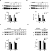
Figure 1.
Figure 1.
Preferential depletion of complex II/SDH subunits in the striatum of HD patients. Levels of Ip and Fp subunits of complex II/SDH were quantified by Western-blot in caudate (A), putamen (B), cerebral cortex (C), and cerebellum (D) of controls and symptomatic HD patients after normalization to total protein content measured in each lane. Neuropathological grade of each HD patient is indicated above the lanes. Note that only the striatum shows marked depletion of SDH constituents. Data represent mean ± SEM *p < 0.05 by unpaired Student's t test.
Figure 2.
Figure 2.
Analysis of the striatal marker calbindin and mitochondrial proteins in controls and HD patients. Levels of calbindin, BclXL, subunit IV of cytochrome oxidase (COX IV), cytochrome c and α-subunit of complex V (α-CV) were analyzed by Western blot in the putamen of control and HD patients. Top, typical Western blots; bottom, results of the image analysis of the blots shown in the top panel. Note that the proteins show no profound modification of expression, whereas SDH constituents are depleted.
Figure 3.
Figure 3.
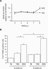
Time course of degeneration in striatal cells expressing Htt171-82Q using lentivirus-mediated gene transfer. Time course of caspase-3 related proteolytic activity was measured using the fluorescent substrate DEVD-AFC. Data are mean ± SEM of two independent experiments performed on three sister cultures per group. Occurrence of actual cell death was counted by FACS after labeling of 10,000 neurons per sister cultures with TUNEL. Note that compared with noninfected cultures (NI) and cells expressing Htt171-19Q (Q19) that show low levels of degeneration, expression of Htt171-82Q produces toxicity that significantly increases from 6 to 8 wk postinfection (p.i.). Data represent mean raw counts ± SEM of two independent experiments (n = 6 wells per condition). *p < 0.0001 using ANOVA followed by post hoc PLSD Fischer test.
Figure 4.
Figure 4.
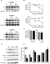
Expression of Htt171-82Q expression of complex II/SDH catalytic subunits. Representative immuno-blots showing Fp, Ip, and α-subunit of complex V (alpha-CV) expression in Htt171-19Q (19Q)- and Htt171-82Q (82Q)-expressing cells after 5, 6, and 8 wk in culture. Levels of expression of Fp and Ip proteins were quantified by Western blot at different time points in control cells (gray stars), and neurons infected with either lenti-Htt171-82Q (filled triangle), or lenti-Htt19Q (filled square). Expression of alpha-CV was also quantified indicating only minor changes. Typical Western blot showing the expression of SDH subunits compared with other mitochondrial proteins at 6 wk postinfection is shown in D and corresponding quantification in E. Note that SDH Ip and Fp show decreased expression, whereas the other proteins are relatively preserved. Data represent mean ± SEM of at least two independent experiments performed on three sister cultures per group for each time point (n = 6 wells per condition). Loss of SDH subunits at 6 wk has been observed in more than four independent experiments. *p < 0.05 by ANOVA and post hoc PLSD Fischer test.
Figure 5.
Figure 5.
Expression of Htt171-82Q induces mitochondrial dysfunction. Dehydrogenase activity of complex II/SDH was measured in Htt177-19Q– and Htt171-82Q–expressing cells at 6 wk postinfection. Activity of complex II (succinate oxidation) was evaluated using the colorimetric substrate INT. Data represent mean ± SEM of two independent experiments with three sister cultures per group. (B) Fluorescence of MitoTracker red in living cells (▪) or after PFA fixation of cultures (▵) as a function of incubation time in presence of the dye. For these methodological controls, fluorescence accumulated in cells was determined using a plate reader. Incubation with the ionophore FCCP (50 μM) produces marked reduction of the accumulation of Mitotracker red, demonstrating that accumulation of the dye is highly sensitive to the loss of mitochondrial membrane potential. (C) Detection of MitoTracker red levels in individual cells after PFA fixation was performed by FACS, showing the typical distribution of fluorescence levels in cells expressing Htt171-82Q (82Q) and Htt171-19Q at 6 wk postinfection. Note the shift to the left of fluorescence distribution in cells expressing Htt171-82Q, indicating reduced fluorescence mean intensity (FMI) in the culture. (D) Histograms representing the quantification of FMI measured in two independent experiments (n = 6 wells per condition). Data are mean ± SEM. *p < 0.05 by ANOVA and post hoc PLSD Fischer test.
Figure 6.
Figure 6.
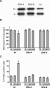
Overexpression of complex II/SDH subunits restores mitochondrial membrane potential and rescues neuronal death induced by Htt171-82Q. (A) Representative Western blots of 30 DIV neurons transduced with SDH encoding lentiviral vectors 2 wk after infection. (B) Overexpression of Fp (SDH-A virus) or Ip (SDH-B virus) suppresses mitochondrial membrane potential alteration induced by Htt171-82Q expression at 6 wk postinfection. (C) Determination of the number of TUNEL-positive cells at 8 wk in culture indicates that both infection with lentiviral vectors coding SDH subunits significantly reduces Htt171-82Q–induced cell death. Data are mean ± SEM of two independent experiments performed on three sister cultures per group (n = 6 wells per condition). *p < 0.05 by ANOVA and post hoc PLSD Fischer test.
Figure 7.
Figure 7.
Overexpression of complex II/SDH subunits protects striatal cell loss rapidly induced by expression of Htt171-82Q under the tetracycline-regulated promoter. The neuroprotective effect of lenti-SDH-A and lenti-SDH-B was tested in a model of degeneration induced by infection with lentiviral vectors coding for Htt-171-82Q under the tetracycline-regulated promoter (TRE). (A) Time course of accumulation of Htt-containing inclusions/aggregates detected by immunofluorescence with the EM48 antibody. Note that inclusion accumulation culminates at 3–4 wk postinfection (p.i.). (B) Inclusions/aggregates in cells expressing TRE-Htt-171-82Q at 4 wk postinfection were detected by immunofluorescence with an anti-ubiquitin antibody. (C) Loss of DARPP32-positive cells at 4 wk postinfection with lenti-TRE-Htt-171-82Q compared with lenti-TRE-Htt171-19Q. The coinfection with lenti-SDH-A and lenti-SDH-B did not markedly modify the density of inclusions and the density of DARPP32 cells. (D) Neuronal nuclei were identified and counted after DNA staining at 4 wk postinfection with lenti-TRE-Htt-171-82Q, lenti-TRE-Htt171-19Q, and lenti-TRE-GFP. When indicated, cultures were coinfected with lenti-SDH-A or lenti-SDH-B. Note that the significant cell loss evidenced in cultures expressing Htt171-82Q is blocked by expression of SDH-A and SDH-B. *p < 0.05 compared with all other groups.

References

    1. Ackrell, B. A. (2000). Progress in understanding structure-function relationships in respiratory chain complex II. FEBS Lett. 466, 1–5. - PubMed
    1. Bae, B. I. et al. (2005). p53 mediates cellular dysfunction and behavioral abnormalities in Huntington's disease. Neuron 47, 29–41. - PubMed
    1. Beal, M. F. (1992). Does impairment of energy metabolism result in excitotoxic neuronal death in neurodegenerative illnesses? Ann. Neurol. 31, 119–130. - PubMed
    1. Benchoua, A., Braudeau, J., Reis, A., Couriaud, C., and Onteniente, B. (2004). Activation of proinflammatory caspases by cathepsin B in focal cerebral ischemia. J. Cereb. Blood Flow Metab. 24, 1272–1279. - PubMed
    1. Benchoua, A., Couriaud, C., Guegan, C., Tartier, L., Couvert, P., Friocourt, G., Chelly, J., Menissier-de Murcia, J., and Onteniente, B. (2002). Active caspase-8 translocates into the nucleus of apoptotic cells to inactivate poly(ADP-ribose) polymerase-2. J. Biol. Chem. 277, 34217–34222. - PubMed

Publication types

MeSH terms