. 2006 Feb 1;26(5):1355-65.
doi: 10.1523/JNEUROSCI.3164-05.2006.

Synaptic Ras GTPase activating protein regulates pattern formation in the trigeminal system of mice

Affiliations

Synaptic Ras GTPase activating protein regulates pattern formation in the trigeminal system of mice

Mark W Barnett et al. J Neurosci. .

Abstract

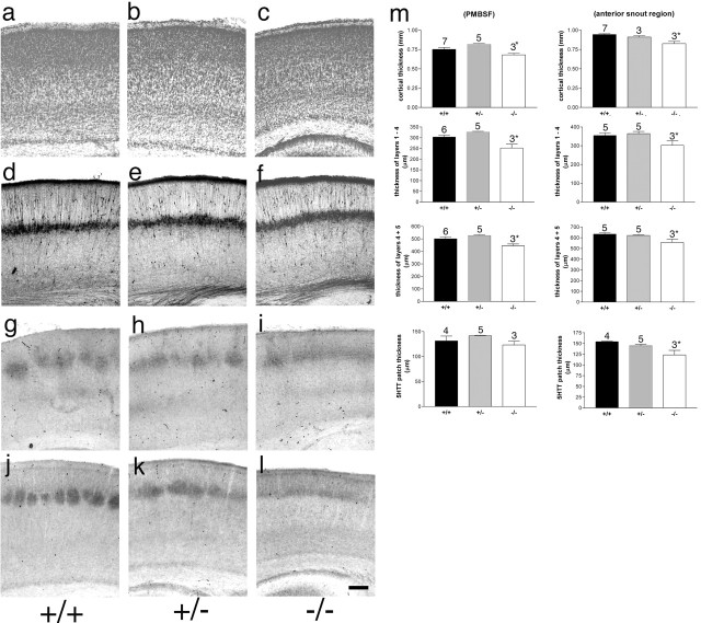
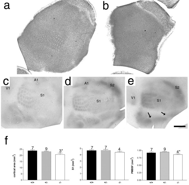
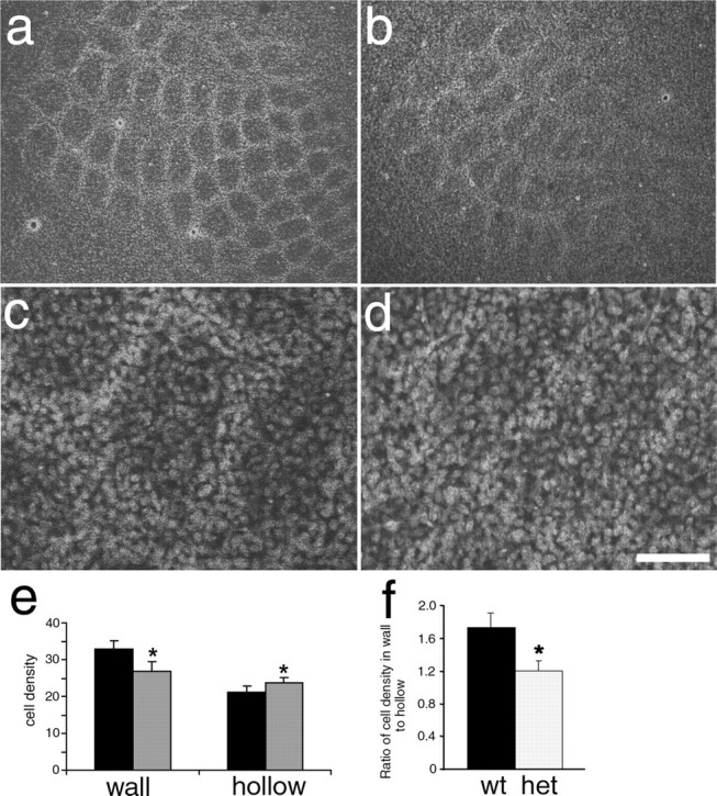
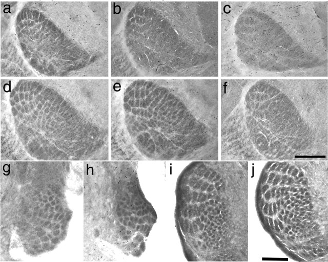
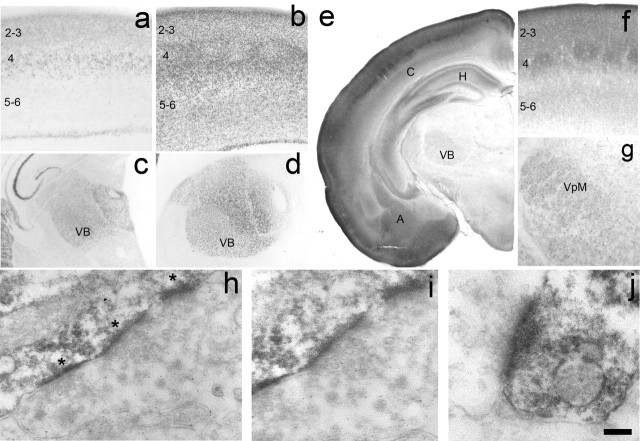
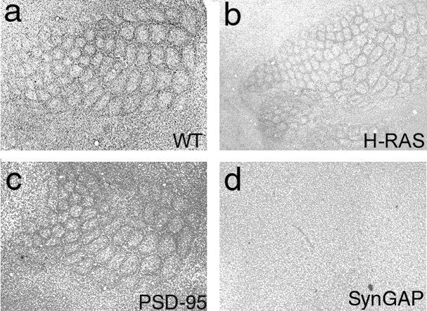
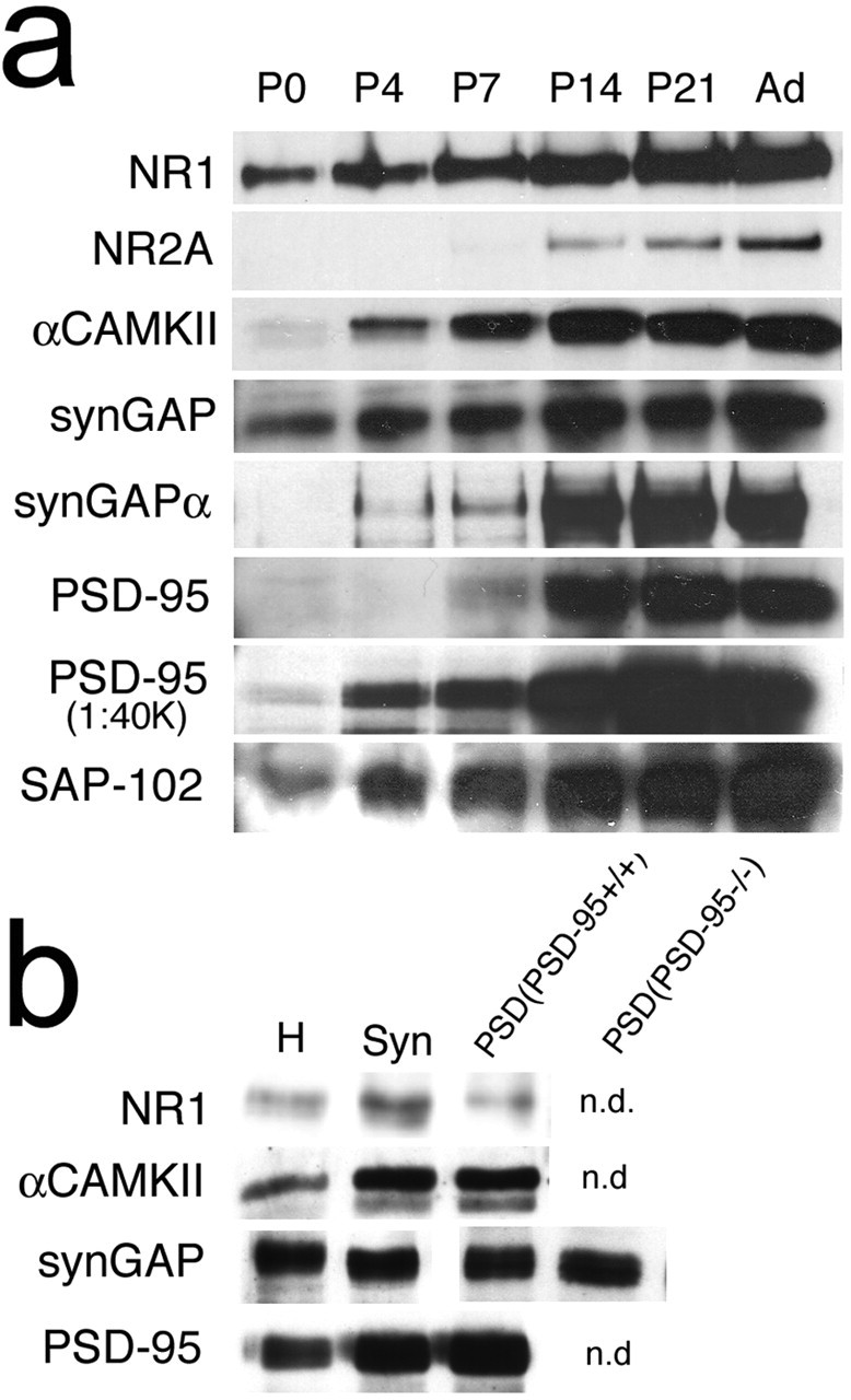
The development of ordered connections or "maps" within the nervous system is a common feature of sensory systems and is crucial for their normal function. NMDA receptors are known to play a key role in the formation of these maps; however, the intracellular signaling pathways that mediate the effects of glutamate are poorly understood. Here, we demonstrate that SynGAP, a synaptic Ras GTPase activating protein, is essential for the anatomical development of whisker-related patterns in the developing somatosensory pathways in rodent forebrain. Mice lacking SynGAP show only partial segregation of barreloids in the thalamus, and thalamocortical axons segregate into rows but do not form whisker-related patches. In cortex, layer 4 cells do not aggregate to form barrels. In Syngap(+/-) animals, barreloids develop normally, and thalamocortical afferents segregate in layer 4, but cell segregation is retarded. SynGAP is not necessary for the development of whisker-related patterns in the brainstem. Immunoelectron microscopy for SynGAP from layer 4 revealed a postsynaptic localization with labeling in developing postsynaptic densities (PSDs). Biochemically, SynGAP associates with the PSD in a PSD-95-independent manner, and Psd-95(-/-) animals develop normal barrels. These data demonstrate an essential role for SynGAP signaling in the activity-dependent development of whisker-related maps selectively in forebrain structures indicating that the intracellular pathways by which NMDA receptor activation mediates map formation differ between brain regions and developmental stage.


Figures

Figure 1.
Figure 1.
Normal cortical lamination in Syngap+/− and Syngap−/− mice. Coronal sections through S1 cortex of P6/7 Syngap+/+ (a, d, g, j), Syngap+/− (b, e, h, k), and Syngap−/− (c, f, i, l) mice stained for Nissl substance (a–c), calretinin (d–f), 5-HTT (g–i), and PKA RIIβ (j–l). No qualitative difference in pattern of staining is visible between genotypes with the exception of the lack of segregation of TCAs seen in the Syngap−/− animals. Quantitative analysis of cortical thickness, radial thickness of TCA terminals, layer 5/6 thickness, and layer 1–4 thickness was also calculated (m). In all cases, there was no significant difference between Syngap+/+ and Syngap+/− animals. There was a significant decrease in Syngap−/− compared with Syngap+/+ and Syngap+/− animals in all parameters measured except 5-HTT terminal zone thickness in PMBSF. A complete numerical account of these data is presented in supplemental Table 1 (available at www.jneurosci.org as supplemental material). Scale bar, 250 μm. Error bars represent SE.
Figure 2.
Figure 2.
Lack of barrel formation in Syngap−/− mice. Nissl staining of flattened sections through layer 4 of P6.5 Syngap+/+ (a) and Syngap−/− (b) mice showing a complete absence of cellular aggregation in Syngap−/− mice. Flattened sections through layer 4 of Syngap+/+ (c), Syngap+/− (d), and Syngap−/− (e) mice were immunostained for 5-HT to reveal the distribution of TCAs. Clear segregation of primary visual (V1), somatosensory (S1), and auditory (A1) as well as secondary somatosensory (S2) cortical areas can be seen in all three genotypes indicating no general defect in TCA pathfinding in Syngap mutants. Within S1, the representation of different body regions (PMBSF and the anterior snout, lower lip, forepaw, and hindpaw representations) are also clearly defined (arrows). However, within the anterior snout, no TCA segregation is visible and, within PMBSF, TCAs can be seen segregating into rows, but patches corresponding to individual whiskers fail to form. Scale bar (in e): a, b, 1 mm; c–e, 800 μm. Quantification of neocortical area, area of S1, and PMBSF in Syngap+/+, Syngap+/−, and Syngap−/− animals revealed a small but significant decrease in neocortical area and area of PMBSF in Syngap−/− animals. The area of S1 was reduced in Syngap−/− animals, although this decrease was not significant. No significant difference between Syngap+/+ and Syngap+/− was seen in any these measurements. In addition, there was no significant difference in the area of individual TCA patches of PMBSF barrels in Syngap+/+ compared with Syngap+/− mice (supplemental Fig. 1, available at www.jneurosci.org as supplemental material). Error bars represent SE.
Figure 3.
Figure 3.
Reduced barrel segregation in Syngap+/− mice. Propidium iodide staining showing neuronal distribution in flattened sections from WT (a, c) and Syngap+/− (b, d) mice showing a clear reduction in the ratio of cells in the barrel wall to barrel hollow in Syngap+/− mice. Cell counts revealed a significant decrease (p < 0.01) in the density of barrel wall neurons and a significant increase (p < 0.01) in the density of barrel hollow neurons in Syngap+/− (gray bars) compared with Syngap+/+ (black bars) animals (e). The barrel wall to hollow ratio is significantly reduced (p < 0.01) from 1.8 in WT mice to 1.2 in Syngap+/− mice (f). The reduction in this ratio appears to be caused by a change in the distribution of cells rather than an overall change in the number of cells (f). Scale bar (in d): a, b, 250 μm; c, d, 70 μm. Error bars represent SE. het, Heterozygotes.
Figure 4.
Figure 4.
Reduced barreloid segregation in Syngap−/− mice. Cytochrome oxidase staining in VpM of thalamus to reveal barreloids in Syngap+/+ (a, d), Syngap+/− (b, e), and Syngap−/− (c, f) mice at P4 (a–c) and P7 (d–f). Barreloids can be clearly seen in all genotypes at both P3/4 and P7; however, segregation is clearly reduced in Syngap−/− mice (n = 5) at both ages relative to Syngap+/+ (n = 8) and Syngap+/− (n = 12) mice, especially in the anterior snout representations. Coronal section through the brainstem trigeminal complex stained for cytochrome oxidase in P3/4 control (g, i; n = 10) and Syngap−/− (h, j; n = 6) mice showing normal barrelette formation in PrV (g, h) and SpI (i, j). Scale bars: (in f) a–f, 350 μm; (in j) g, h, 225 μm; (in j) i, j, 250 μm.
Figure 5.
Figure 5.
SynGAP expression in the developing cortex and thalamus. Histochemistry for β-galactosidase to reveal the expression profile of Syngap in the S1 (a, b) and thalamus (c, d) at P4 (a, c) and P8 (b, d). For a complete developmental series, see supplemental Figure 2 (available at www.jneurosci.org as supplemental material). In cortex, β-gal is first expressed at P0 in layer 4 of the developing cortical plate. By P4, staining can be seen throughout the supragranular layers as well as layer 4 and the upper region of layer 5. It is also present in a thin strip of cells located at the bottom of the cortical plate (a). By P8, staining can be seen throughout the cortical plate, and the barrels are clearly distinguished in layer 4 (c). By adulthood, staining has been dramatically reduced and can only be seen at the layer 4/5 border and in layer 1. A similar developmental profile can be seen in the VB and throughout the dorsal thalamus with strong staining visible at P4 (c) and P8 (d) and reduced expression in adults. Immunohistochemical localization of SynGAP protein at P7 (e–g) is in good agreement with β-gal localization. Low-power images show high levels of SynGAP in the cortex (C), hippocampus (H), and amygdaloid complex (A) with lower levels in the VB of the thalamus. In S1 (f), SynGAP levels are highest in layer 4, and the supragranular layers and barrels can be clearly seen. Expression is just starting to appear in the infragranular layers, although a dense band of label can be seen at the layer 4/5 border. The immunohistochemical localization therefore is in good agreement with the large increase in X-Gal staining between P4 and P7. In thalamus, SynGAP is expressed throughout the VB. Electron microscopy analysis of SynGAP expression in layer 4 at P14 reveals a postsynaptic localization for SynGAP (h–j). DAB reaction product can be clearly seen as dark labeling in the PSD (asterisks) opposing presynaptic terminals containing synaptic vesicles. In h, three PSDs in tandem on the shaft of a dendrite are clearly visible. Higher magnification of the middle synapse in h shows clear amalgamation of presynaptic vesicles abutting the SynGAP-positive PSD. Clear presynaptic vesicles are also seen in j. Reaction product was never seen in axons or axon terminals. Scale bar (in j): a, b, 200 μm; c, d, 400 μm; e, 775 μm; f, 150 μm; g, 275 μm; h, 250 nm; i, 175 nm; j, 100 nm.
Figure 6.
Figure 6.
Normal barrel formation in mice lacking PSD-95 and H-Ras. Nissl staining in flattened sections through layer 4 of WT (a), H-Ras (b), PSD-95 (c), and SynGAP (d) mutant mice.
Figure 7.
Figure 7.
SynGAP still associates with the PSD in Psd-95−/− mice. Western blotting for PSD components in homogenates of S1 cortex reveals a dramatic increase in PSD components during the first postnatal week (a). b, Western blotting for NR1, CaMKII, PSD-95, and SynGAP (Syn) in homogenates, synaptosomes, and PSDs isolate from S1 of P7 WT mice as well as SynGAP expression in the PSD of P7 Psd-95−/− mice. n.d., Not done.

References

    1. Adams JP, Sweatt JD (2002). Molecular psychology: roles for the ERK MAP kinase cascade in memory. Annu Rev Pharmacol Toxicol 42:135–163. - PubMed
    1. Barnett MW, Old RW, Jones EA (1998). Neural induction and patterning by fibroblast growth factor, notochord and somite tissue in Xenopus. Dev Growth Differ 40:47–57. - PubMed
    1. Berkeley JL, Levey AI (2003). Cell-specific extracellular signal-regulated kinase activation by multiple G protein-coupled receptor families in hippocampus. Mol Pharmacol 63:128–135. - PubMed
    1. Bozon B, Kelly A, Josselyn SA, Silva AJ, Davis S, Laroche S (2003). MAPK, CREB and zif268 are all required for the consolidation of recognition memory. Philos Trans R Soc Lond B Biol Sci 358:805–814. - PMC - PubMed
    1. Chen HJ, Rojas-Soto M, Oguni A, Kennedy MB (1998). A synaptic Ras-GTPase activating protein (p135 synGAP) inhibited by CaM kinase II. Neuron 20:895–904. - PubMed

Publication types

MeSH terms

Substances

LinkOut - more resources