. 2006 Feb 1;26(5):1366-77.
doi: 10.1523/JNEUROSCI.3889-05.2006.

Physiological temperatures reduce the rate of vesicle pool depletion and short-term depression via an acceleration of vesicle recruitment

Affiliations

Physiological temperatures reduce the rate of vesicle pool depletion and short-term depression via an acceleration of vesicle recruitment

Christopher Kushmerick et al. J Neurosci. .

Abstract

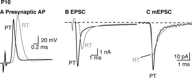
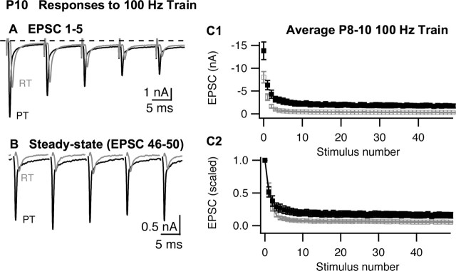
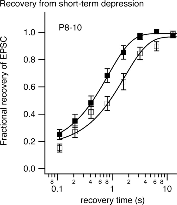
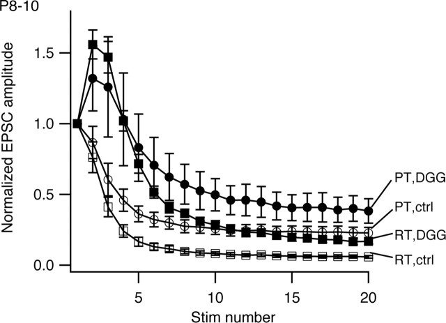
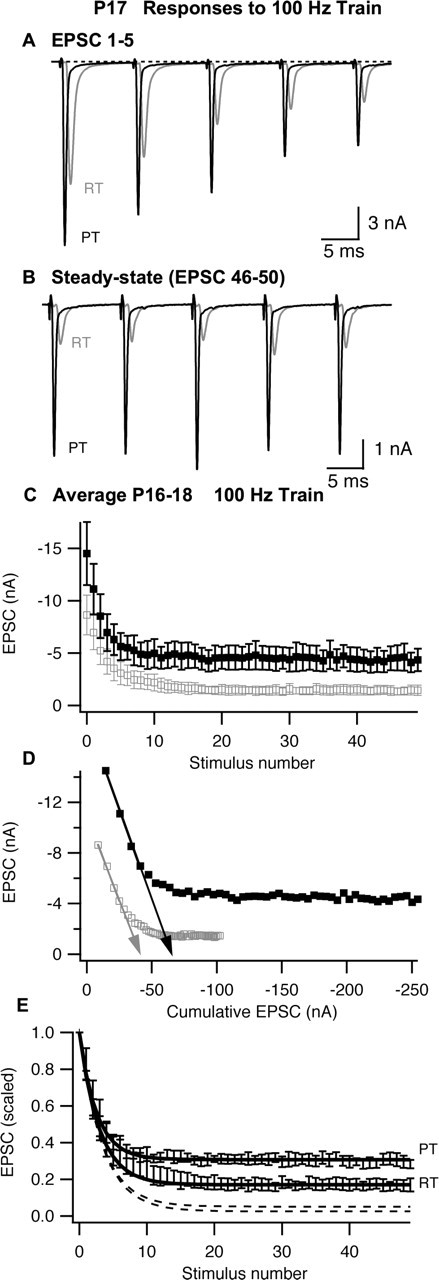
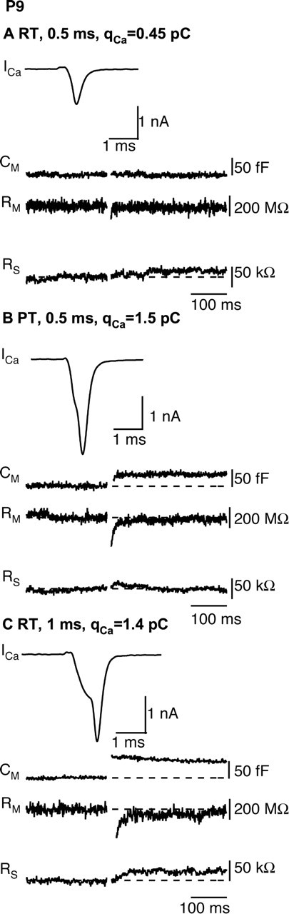
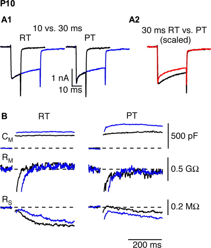
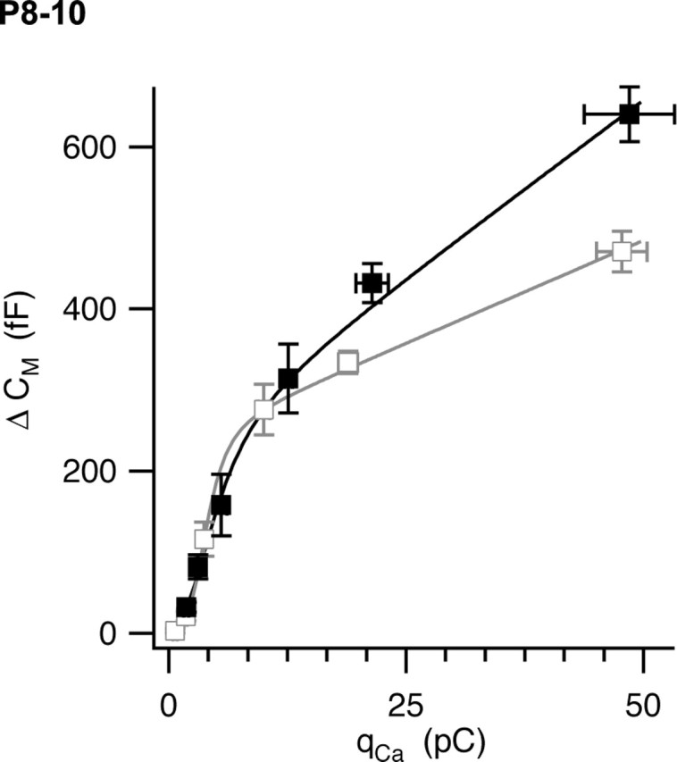
The timing and strength of synaptic transmission is profoundly dependent on temperature. However, the temperature dependence of the multiple mechanisms that contribute to short-term synaptic plasticity is poorly understood. Here, we use voltage-clamp recordings to quantify the temperature dependence of exocytosis at the calyx of Held synapse. EPSC and miniature EPSC amplitudes were larger at physiological temperature, but quantal content during low-frequency (0.05 Hz) stimulation was constant after temperature jumps from 22-24 degrees C to 35-37 degrees C. The initial degree of EPSC depression during 100 Hz stimuli trains was unchanged with temperature, as were estimates of release probability and vesicle pool size. In contrast, physiological temperatures dramatically relieved depression measured after 40 stimuli at 100 Hz by increasing twofold the rate of recovery from depression. Presynaptic calyx recordings revealed that physiological temperature increased capacitance jumps resulting from 0.5 and 1 ms depolarizations by increasing Ca2+ influx. When Ca2+ entry was equalized at the two temperatures, exocytosis exhibited little temperature dependence for brief depolarizations. However, in response to longer depolarizations, raising temperature increased a slow phase of exocytosis, without affecting overall Ca2+ entry or the size of the readily releasable pool of vesicles. Higher temperatures also increased the rate of presynaptic Ca2+ current inactivation; nevertheless, the degree of steady-state EPSC depression was greatly reduced. Our results thus suggest that changes in steady-state EPSCs during stimulus trains at physiological temperature reflect larger quantal amplitudes and faster refilling of synaptic vesicle pools, leading to reduced short-term depression during prolonged high-frequency firing.


Figures

Figure 1.
Figure 1.
The presynaptic action potential (A), EPSC (B), and average mEPSC (C) recorded in the MTNB from a P10 animal at RT (gray) or PT (black). The data shown in B and C were recorded from the same cell. The mEPSCs shown in C are the average of 74 and 231 miniature events recorded at room temperature or physiological temperature, respectively.
Figure 2.
Figure 2.
Synaptic depression recorded in P8–P10 MNTB neurons during stimulation of the calyx of Held with 100 Hz trains. A, B, The first five EPSCs (A) or last five EPSCs (B) recorded from a P10 MNTB neuron during a train of 50 stimuli at RT (gray) or PT (black). In B, stimulus artifacts were removed for clarity. C, Summary (n = 9 cells) of EPSC amplitudes (C1) or scaled amplitudes (C2) during 100 Hz stimulus train at room temperature (open symbols; gray) or physiological temperature (filled symbols; black). The error bars represent the SEM. In C2, EPSC amplitudes for each cell were scaled to the first EPSC in the respective train, and then mean and SEM of the normalized data set was calculated.
Figure 3.
Figure 3.
Recovery from synaptic depression recorded in P8–P10 MNTB neurons at room temperature (open symbols) or physiological temperature (filled symbols). A conditioning train of 20 stimuli at 100 Hz was delivered followed by a variable recovery time before a test stimuli was delivered. The interval between successive conditioning trains was 20 s. Fractional recovery was calculated as follows: (AAss)/(AoAss), where A is the amplitude of the test EPSC after a given recovery time, Ao is the amplitude of the first EPSC in the conditioning train, and Ass is the steady-state EPSC amplitude at the end of the conditioning train. The solid curves represent exponential fits to the data. Error bars represent SEM.
Figure 4.
Figure 4.
Protecting AMPA receptors from desensitization with 4 mm γ-DGG does not antagonize effects of temperature on steady-state depression in MNTB from P8–P10 animals. The mean normalized EPSC amplitudes (n = 6 cells) recorded in either normal aCSF (open symbols; ctrl) or in the presence of 4 mm γ-DGG (filled symbols; DGG) and at RT (squares) or PT (circles) are shown. Error bars represent SEM. Stim, Stimulus.
Figure 5.
Figure 5.
Synaptic depression recorded in P16–P18 MNTB neurons during stimulation of the calyx of Held with 100 Hz trains. A, B, The first five EPSCs (A) or last five EPSCs (B) recorded from a P17 MNTB neuron during a train of 50 stimuli at RT or PT. In B, stimulus artifacts were removed for clarity. C, Summary (n = 5 cells) of EPSC amplitudes. D, Determination of vesicle pool size and release probability by linear extrapolation (arrows) of EPSC amplitude versus cumulative EPSC amplitude. E, Normalized EPSC amplitudes. The dashed lines represent model predictions (see Materials and Methods) based on vesicle refilling rates obtained from measurements of recovery from synaptic depression (see Fig. 3) at RT or PT, and the solid lines represent model predictions when the recovery rate was adjusted to fit observed steady-state depression. EPSC amplitudes for each cell were scaled to the first EPSC in the respective train, and the mean and SEM of the normalized data set were calculated. In C and D, the open gray symbols represent data recorded at room temperature, and filled black symbols represent data recorded at physiological temperature; in E, symbols were omitted for clarity. Error bars represent SEM.
Figure 6.
Figure 6.
Temperature dependence of exocytosis during brief depolarizations. A, B, A 0.5 ms depolarization to 0 mV evokes a capacitance jump at physiological temperature but not at room temperature. Voltage-clamp recordings of ICa, Cm, membrane resistance (Rm), and series resistance (RS) in response to 0.5 ms depolarization to 0 mV at room temperature (A) or physiological temperature (B) are shown. C, A 1.0 ms depolarization at room temperature generates robust ICa and Cm jump. The data in A–C were obtained from the same calyx of Held.
Figure 7.
Figure 7.
The late phase of exocytosis during prolonged depolarizations is temperature sensitive. The calyx was depolarized for 10 ms (black) or 30 ms (blue) from −80 to 0 mV at room temperature (left) or physiological temperature (right). A, Presynaptic Ca2+ currents. A2, Ca2+ current inactivation was more pronounced at PT (red) than at RT (black), as seen after scaling the currents by their peak amplitudes. The activation kinetics of the Ca2+ current was faster at PT. B, Changes in Cm, membrane resistance (Rm), and series resistance (RS) in response to 10 and 30 ms depolarizations at RT and PT {depolarizing step pulses for 10 ms (black) or 30 ms (blue) as shown in A1}.
Figure 8.
Figure 8.
Ca2+–exocytosis coupling in the calyx of Held at room temperature (open symbols; gray) and physiological temperature (filled symbols; black). The calyx was depolarized under voltage clamp for variable times ranging from 0.5 to 30 ms. Resulting capacitance jumps (ΔCm) were plotted against the integral of the Ca2+ current (qCa). Error bars represent SEM (n = 9–25 measurements per point). The solid curves represent fits of a model equation described in Results.

References

    1. Allen C, Stevens CF (1994). An evaluation of causes for unreliability of synaptic transmission. Proc Natl Acad Sci USA 91:10380–10383. - PMC - PubMed
    1. Augustine G (1990). Regulation of transmitter release at the squid giant synapse by presynaptic delayed rectifier potassium current. J Physiol (Lond) 431:343–364. - PMC - PubMed
    1. Awatramani GB, Turecek R, Trussell LO (2004). Inhibitory control at a synaptic relay. J Neurosci 24:2643–2647. - PMC - PubMed
    1. Billups B, Graham BP, Wong AY, Forsythe ID (2005). Unmasking group III metabotropic glutamate autoreceptor function at excitatory synapses in the rat CNS. J Physiol (Lond) 565:885–896. - PMC - PubMed
    1. Bollmann JH, Sakmann B, Borst JG (2000). Calcium sensitivity of glutamate release in a calyx-type terminal. Science 289:953–957. - PubMed

Publication types

Substances

LinkOut - more resources