. 2006 Feb 1;26(5):1407-17.
doi: 10.1523/JNEUROSCI.3463-05.2006.

The dopaminergic midbrain participates in human episodic memory formation: evidence from genetic imaging

Affiliations

The dopaminergic midbrain participates in human episodic memory formation: evidence from genetic imaging

Björn H Schott et al. J Neurosci. .

Abstract

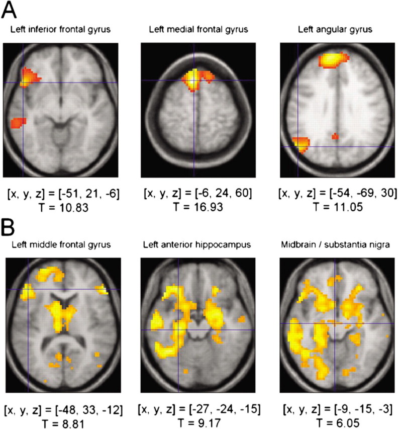
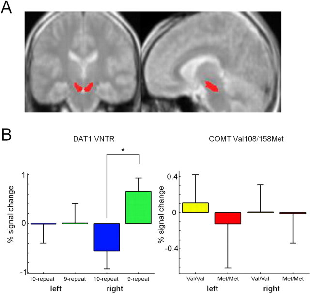
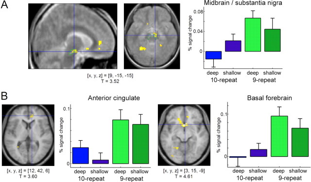
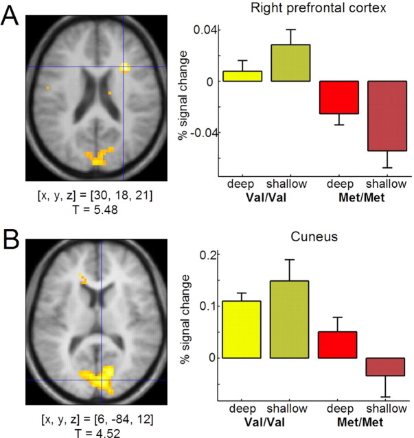
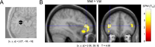
Recent data from animal studies raise the possibility that dopaminergic neuromodulation promotes the encoding of novel stimuli. We investigated a possible role for the dopaminergic midbrain in human episodic memory by measuring how polymorphisms in dopamine clearance pathways affect encoding-related brain activity (functional magnetic resonance imaging) in an episodic memory task. In 51 young, healthy adults, successful episodic encoding was associated with activation of the substantia nigra. This midbrain activation was modulated by a functional variable number of tandem repeat (VNTR) polymorphism in the dopamine transporter (DAT1) gene. Despite no differences in memory performance between genotype groups, carriers of the (low expressing) 9-repeat allele of the DAT1 VNTR showed relatively higher midbrain activation when compared with subjects homozygous for the 10-repeat allele, who express DAT1 at higher levels. The catechol-O-methyl transferase (COMT) Val108/158Met polymorphism, which is known to modulate enzyme activity, affected encoding-related activity in the right prefrontal cortex (PFC) and in occipital brain regions but not in the midbrain. Moreover, subjects homozygous for the (low activity) Met allele showed stronger functional coupling between the PFC and the hippocampus during encoding. Our finding that genetic variations in the dopamine clearance pathways affect encoding-related activation patterns in midbrain and PFC provides strong support for a role of dopaminergic neuromodulation in human episodic memory formation. It also supports the hypothesis of anatomically and functionally distinct roles for DAT1 and COMT in dopamine metabolism, with DAT1 modulating rapid, phasic midbrain activity and COMT being particularly involved in prefrontal dopamine clearance.


Figures

Figure 1.
Figure 1.
Activations related to level of processing and subsequent memory in the entire cohort of 51 subjects. A, Level of processing effect. Regions in which HRF distinguishes deeply studied items from shallowly studied items include the left inferior frontal gyrus (left), the bilateral medial prefrontal cortex (middle), and the angular gyrus (right). B, Overall group effects of subsequent memory, regardless of genotype. Activations in the left dorsolateral prefrontal cortex (left), in the anterior hippocampus (middle), and in the midbrain (right) are greater for subsequently remembered compared with subsequently forgotten items. Activations above a threshold of T > 5.27 (p < 0.05, full-volume corrected; extent threshold, k = 10 voxels) are displayed. [x, y, z], Voxel coordinates in MNI (Montreal Neurological Institute) reference space.
Figure 2.
Figure 2.
Effects of DAT1 and COMT genotypes on memory-related midbrain activations. A, ROIs encompassing the left and right substantia nigra and the ventral tegmental area. The ROIs were segmented manually from an average of five normalized MT images. B, Results of the ROI analyses in the left and right midbrain for the DAT1 VNTR (left) and the COMT Val108/158Met polymorphism (right). The 9-repeat carriers of the DAT1 VNTR showed significantly higher activation of the right midbrain during successful episodic memory formation. *p < 0.05, partial volume corrected.
Figure 3.
Figure 3.
Effects of the DAT1 3′ VNTR polymorphism on memory-related brain activity. A, The 9-repeat carriers show significantly higher memory-related midbrain activations than 10-repeat homozygous subjects. To verify the localization of the effect, the cluster of activation has been superimposed onto the normalized average of five MT images, on which the substantia nigra can be clearly distinguished from surrounding structures (Eckert et al., 2004). The substantia nigra has been segmented for illustrative purposes (green). B, The 9-repeat carriers also show significantly higher activations in the anterior cingulate (left) and in the basal forebrain (right) for subsequently remembered compared with subsequently forgotten items. Activations above a threshold of T > 2.72 (p < 0.005, uncorrected; extent threshold, k = 10 voxels) are displayed. Bar plots depict peak percentages of signal change of the canonical HRFs (SPM betas for subsequently remembered − subsequently forgotten items; scaled to the HRF) and their SEs for both groups, separately for the deep and shallow study conditions. [x, y, z], Voxel coordinates in MNI reference space.
Figure 4.
Figure 4.
Effects of COMT Val108/158Met polymorphism on memory-related brain activity. Compared with Met homozygous subjects, Val homozygous subjects show increased memory-related activations of the right prefrontal cortex (A) and of the cuneus, extending into the primary visual cortex (B). Activations above a threshold of T > 2.72 (p < 0.005, uncorrected; extent threshold, k = 10 voxels) are displayed. Bar plots display the same information as in Figure 3. [x, y, z], Voxel coordinates in MNI reference space.
Figure 5.
Figure 5.
Effects of COMT Val108/158Met polymorphism on functional coupling of the prefrontal cortex and the hippocampus. A, A representative volume of interest from a single subject. Volumes of interest were spheres with a radius of 6 mm and contained up to 32 voxels. B, Compared with Val homozygous subjects, Met/Met carriers showed significantly stronger functional coupling between the left hippocampus and both the left and right prefrontal cortex. [x, y, z], Voxel coordinates in MNI reference space.
Figure 6.
Figure 6.
Expression of DAT1 in the human hippocampus. A, In the CA3 region, a fine network of DAT1-immunoreactive fibers could be observed. B, In the subiculum, a small subset of DAT1-immunoreactive neurons stood out. C, Western blotting revealed a DAT1-immunoreactive band at ∼85 kDa in both the human striatum and hippocampus. HC, Hippocampus; Str, striatum.

References

    1. Aalto S, Bruck A, Laine M, Nagren K, Rinne JO (2005). Frontal and temporal dopamine release during working memory and attention tasks in healthy humans: a positron emission tomography study using the high-affinity dopamine D2 receptor ligand [11C]FLB 457. J Neurosci 25:2471–2477. - PMC - PubMed
    1. Addis DR, McIntosh AR, Moscovitch M, Crawley AP, McAndrews MP (2004). Characterizing spatial and temporal features of autobiographical memory retrieval networks: a partial least squares approach. NeuroImage 23:1460–1471. - PubMed
    1. Akil M, Kolachana BS, Rothmond DA, Hyde TM, Weinberger DR, Kleinman JE (2003). Catechol-O-methyltransferase genotype and dopamine regulation in the human brain. J Neurosci 23:2008–2013. - PMC - PubMed
    1. Bach ME, Barad M, Son H, Zhuo M, Lu YF, Shih R, Mansuy I, Hawkins RD, Kandel ER (1999). Age-related defects in spatial memory are correlated with defects in the late phase of hippocampal long-term potentiation in vitro and are attenuated by drugs that enhance the cAMP signaling pathway. Proc Natl Acad Sci USA 96:5280–5285. - PMC - PubMed
    1. Barcelo F, Suwazono S, Knight RT (2000). Prefrontal modulation of visual processing in humans. Nat Neurosci 3:399–403. - PubMed

Publication types

MeSH terms

Substances