. 2006 Feb 1;26(5):1439-47.
doi: 10.1523/JNEUROSCI.2219-05.2006.

Proliferation and death of oligodendrocytes and myelin proteins are differentially regulated in male and female rodents

Affiliations

Proliferation and death of oligodendrocytes and myelin proteins are differentially regulated in male and female rodents

Mirela Cerghet et al. J Neurosci. .

Abstract

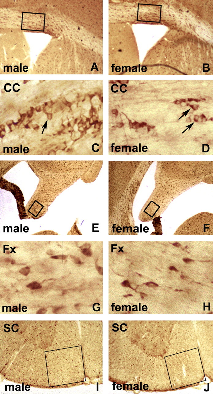
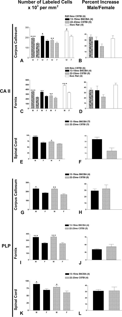
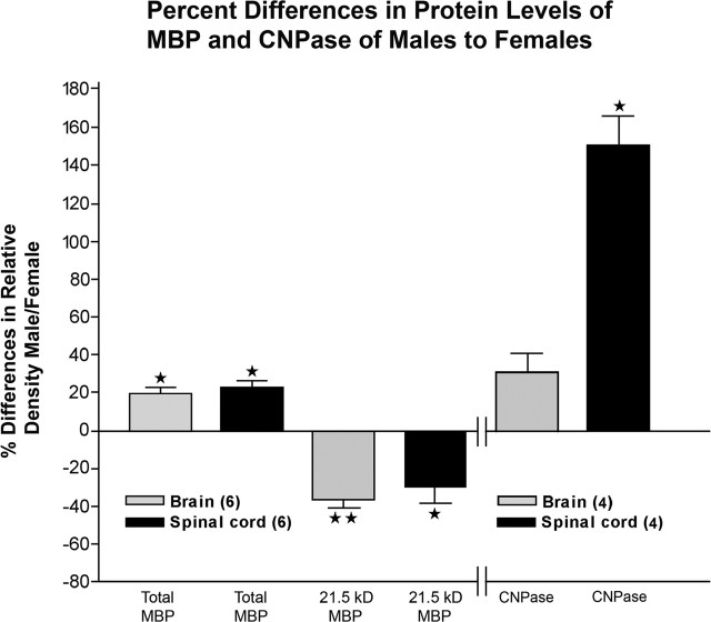
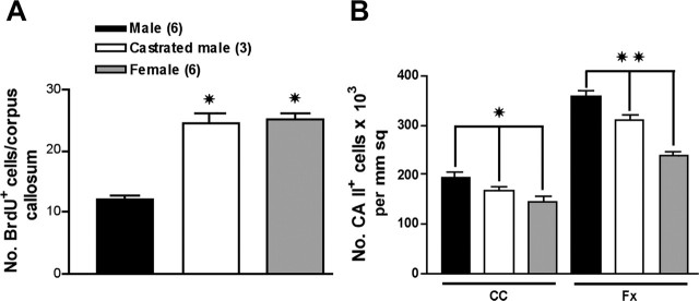
Sexual dimorphism of neurons and astrocytes has been demonstrated in different centers of the brain, but sexual dimorphism of oligodendrocytes and myelin has not been examined. We show, using immunocytochemistry and in situ hybridization, that the density of oligodendrocytes in corpus callosum, fornix, and spinal cord is 20-40% greater in males compared with females. These differences are present in young and aged rodents and are independent of strain and species. Proteolipid protein and carbonic anhydrase-II transcripts, measured by real-time PCR, are approximately two to three times greater in males. Myelin basic protein and 2', 3'-cyclic nucleotide 3'-phosphodiesterase, measured by Western blots, are 20-160% greater in males compared with females. Surprisingly, both generation of new glia and apoptosis of glia, including oligodendrocytes, are approximately two times greater in female corpus callosum. These results indicate that the lifespan of oligodendrocytes is shorter in females than in males. Castration of males produces a female phenotype characterized by fewer oligodendrocytes and increased generation of new glia. These findings indicate that exogenous androgens differentially affect the lifespan of male and female oligodendrocytes, and they can override the endogenous production of neurosteroids. The data imply that turnover of myelin is greater in females than in males. Mu-calpain, a protease upregulated in degeneration of myelin, is dramatically increased at both transcriptional and translational levels in females compared with males. These morphological, molecular, and biochemical data show surprisingly large differences in turnover of oligodendrocytes and myelin between sexes. We discuss the potential significance of these differences to multiple sclerosis, a sexually dimorphic disease, whose progression is altered by exogenous hormones.


Figures

Figure 1.
Figure 1.
Carbonic anhydrase II immunoperoxidase staining of vibratomed sections of corpora callosa (A–D), fornices (E–H), and spinal cords (I, J) of 22-month-old C57BL/6J mice. Low magnification of male (A) and female (B) corpora callosa (CC) at striatal levels shows boxed areas used for quantification of CA II cells. Higher magnification illustrates immunostained Olg cytoplasm (arrows) in males (C) and females (D). Olgs were counted as positive that have unstained nuclei surrounded by cytoplasmic staining. Olgs are typically arranged in rows, interspersed with astrocytes. Low magnification of male (E) and female (F) fornices (Fx) shows boxed areas used for quantification. Higher magnification shows cytoplasmic Olg immunostaining in male (G) and female (H). Low magnification of spinal cord (SC) ventral funiculus adjacent to ventral fissure shows boxed areas used for quantification in male (I) and female (J) mice.
Figure 2.
Figure 2.
In situ hybridization staining using digoxigenin-labeled PLP/DM20 cDNAs of corpora callosa (A–D), fornices (E–H), and cervical spinal cord (I, J) at the striatal level of 22-month-old C57BL/6J mice. Low magnification of male (A) and female (B) corpora callosa (CC) at striatal levels shows boxed areas used for quantification. Higher magnification of male (C) and female (D) corpora callosa shows staining of Olg cytoplasm (arrows). Olgs were counted as positive that have perinuclear cytoplasm staining denoting the presence of PLP mRNA. Low magnification of male (E) and female (F) fornices (Fx) showing boxed areas used for quantification. Higher magnification shows cytoplasmic Olg immunostaining in males (G) and females (H). Low magnification of spinal cord (SC) ventral funiculus shows boxed area used for quantification in male (I) and female (J) mice.
Figure 3.
Figure 3.
Quantification of CA II-immunostained cells located in boxes shown for corpora callosa (A), for fornices (C), and for ventral columns of spinal cord (E) and cells expressing PLP message in corpora callosa (G), in fornices (I), and in ventral columns of spinal cords (K). Percentage increases in CA II cells in corpora callosa (B), in fornices (D), and in ventral columns of spinal cord (F) and cells expressing PLP message in corpora callosa (H), in fornices (J), and in ventral columns of spinal cord (L). Values represent mean ± SEM of three to four sections per animal; number of animals counted at each age are in parentheses next to rodent strain. Two-tailed t test comparison of male with female, assuming equal variance was used; *p < 0.05; **p < 0.001; ***p < 0.0001.
Figure 4.
Figure 4.
Percentage differences in relative densities of the four MBP isoforms in brain and spinal cord and CNPase in brains of 8- to 9-month-old C57BL/6 mice. The ratio of relative density of total MBP (sum of values for all 4 isoforms) and β-actin was calculated, and percentage differences in males were compared with females. The values for 21.5 kDa isoforms were separately expressed because the ratio of 21.5 kDa is greater in females than in males. Values are the mean ± SEM of six male and six female pairs. The percentage difference in relative density of CNPase in the brain and spinal cord of four males compared with four females was similarly calculated as for MBP. One-sample t test was used; *p < 0.05; **p < 0.001.
Figure 5.
Figure 5.
Generation of glial cells in corpus callosum. Representative low-magnification pictures at the level of the corpus callosum of 6-week-old male (A) and female (B) CDIRS rats immunolabeled for BrdU. The large number of BrdU+ cells in the subventricular zone of the female is a consistent finding at all ages. Higher magnification of BrdU+ cells in corpora callosa of male (C) and female (D) rats. One injection of BrdU was given daily for 3 d, and the animals were killed 1 week after the first injection. Double immunostaining of 9-month-old C57BL/6J corpus callosum for BrdU/GFAP (E), BrdU/CA II (F), or elimination of primary antibody (G). GFAP+ and CA II+ cells (arrows) are visualized in brown, and their BrdU+ nuclei are visualized in black. The percentage of Olgs and astrocytes that are BrdU+ is similar in males and females (H).
Figure 6.
Figure 6.
Total number of BrdU+ glia in corpora callosa at different ages and in different rodent strains (A). The number of BrdU+ cells is the sum of both sides of the corpora callosa at the level of the striatum. Rats at 6 weeks have nearly 10 times the number of BrdU+ cells as the mouse. The percentage increase in female rodents is approximately two times greater at all four ages (B) in mice and rats. Values represent mean ± SEM of BrdU+ cells in corpora callosa from six to eight sections per animal, and number of animals at each age is in parentheses. Two-tailed t test assuming equal variances was used to compare male with female for each strain; *p < 0.05; **p < 0.0001.
Figure 7.
Figure 7.
Examples of cleaved caspase-3+ cells in corpus callosum visualized with DAB (A, B). An apoptotic glia in an advanced stage of degeneration shows cleaved caspase-3 immunostaining in the nucleus (arrow); an apoptotic cell in an early stage of degeneration shows cleaved caspase-3 immunostaining in the cytoplasm (arrowhead) and a cell with wrinkled membrane and light vacuoles inside (asterisk); elimination of primary antibody shows no staining (C). Double immunostaining with the A007 Olg-specific antibody (D) and cleaved caspase-3 antibody (E) shows that Olgs are apoptotic (F). Scale bars, 5 μm.
Figure 8.
Figure 8.
Western blot of μ-calpain zymogen (110 kDa) and its cleaved form (80 kDa) in brains of three pairs of C57BL/6J mice.
Figure 9.
Figure 9.
The number of BrdU+ cells in corpora callosa of castrated males matches the number in females at 9 months (A). Males were castrated at 6 months and injected daily with BrdU over 3 d beginning 1 week before death at 9 months. The numbers of BrdU+ cells in unoperated males and females is the same data as that shown in the 9 month study (Fig. 6). The number of newly generated BrdU+ cells in castrated males is two times compared with wild-type males. Values represent mean ± SEM of BrdU+ cells in six sections per animal of 9-month-old C57BL/6J. Two-tailed t test assuming equal variances was used to compare male with castrated male and female; *p < 0.001. The number of CA II+ cells in corpus callosum and fornix of castrated mice is decreased compared with unoperated males (B). The decrease in CA II+ Olgs is approximately halfway between the values for unoperated males and females. Values represent mean ± SEM of CA II+ cells. One-way ANOVA was used to compare each group within the corpus callosum (CC) and fornix (Fx); *p < 0.01; **p < 0.0001.

References

    1. Alonso G (2000). Prolonged corticosterone treatment of adult rats inhibits the proliferation of oligodendrocyte progenitors present throughout white and gray matter regions of the brain. Glia 31:219–231. - PubMed
    1. Andrade JP, Madeira MD, Paula-Barbosa MM (2000). Sexual dimorphism in the subiculum of the rat hippocampal formation. Brain Res 875:125–137. - PubMed
    1. Bansal R, Stefansson K, Pfeiffer SE (1992). Proligodendroblast antigen (POA), a developmental antigen expressed by AOO7/O4-positive oligodendrocyte progenitors prior to the appearance of sulfatide and galactocerebroside. J Neurochem 58:2221–2229. - PubMed
    1. Barrera A, Jimenez L, Gonzalez GM, Montiel J, Aboitiz F (2001). Dendritic structure of single hippocampal neurons according to sex and hemisphere of origin in middle-aged and elderly human subjects. Brain Res 906:31–37. - PubMed
    1. Bebo BF Jr, Vandenbark AA, Offner H (1996). Male SJL mice do not relapse after induction of EAE with PLP 139–151. J Neurosci Res 45:680–689. - PubMed

Publication types

MeSH terms