. 2006 Feb 1;26(5):1486-98.
doi: 10.1523/JNEUROSCI.4749-05.2006.

Coordination and modulation of locomotion pattern generators in Drosophila larvae: effects of altered biogenic amine levels by the tyramine beta hydroxlyase mutation

Affiliations

Coordination and modulation of locomotion pattern generators in Drosophila larvae: effects of altered biogenic amine levels by the tyramine beta hydroxlyase mutation

Lyle E Fox et al. J Neurosci. .

Abstract

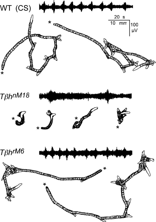
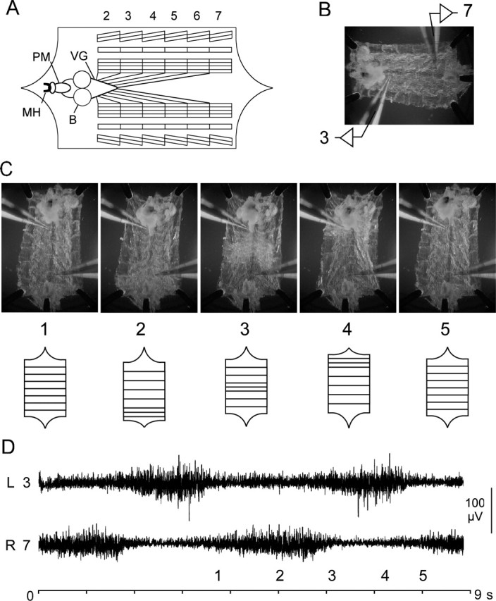
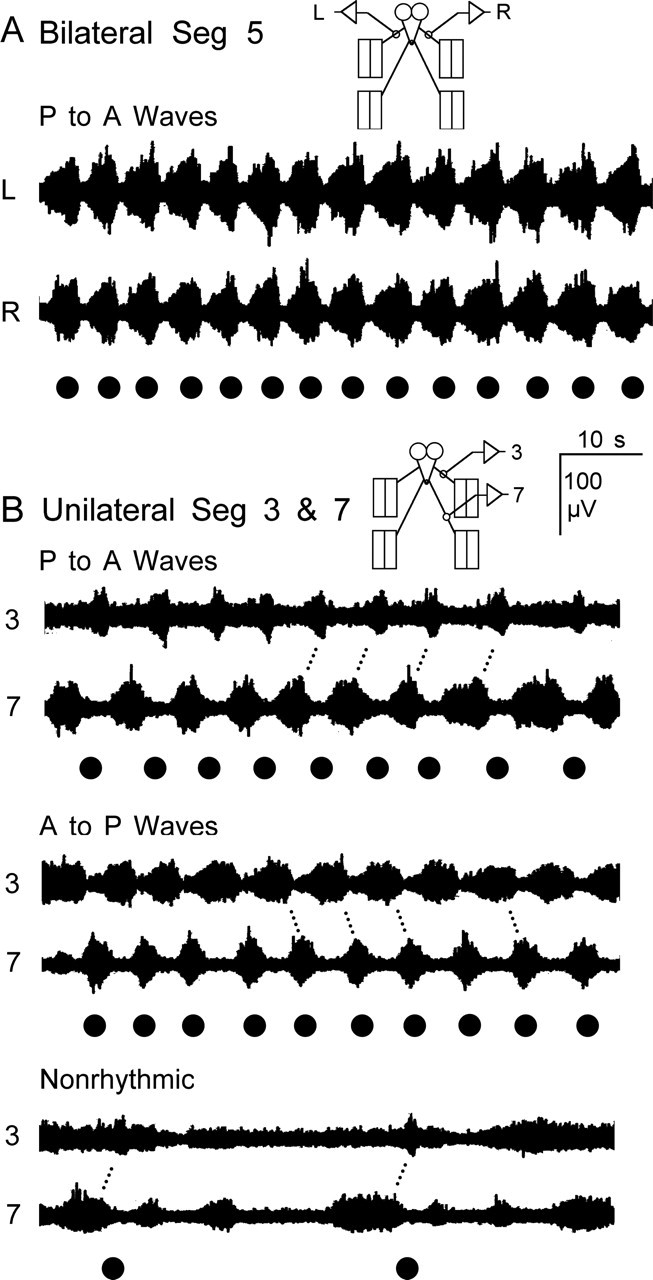
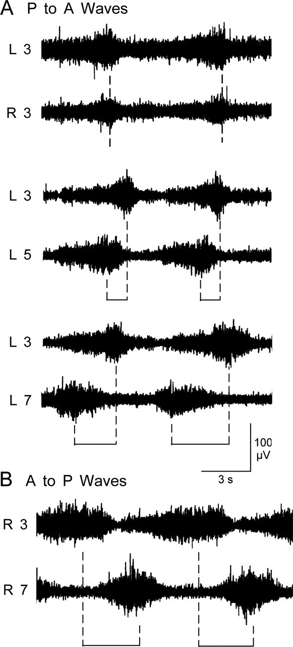
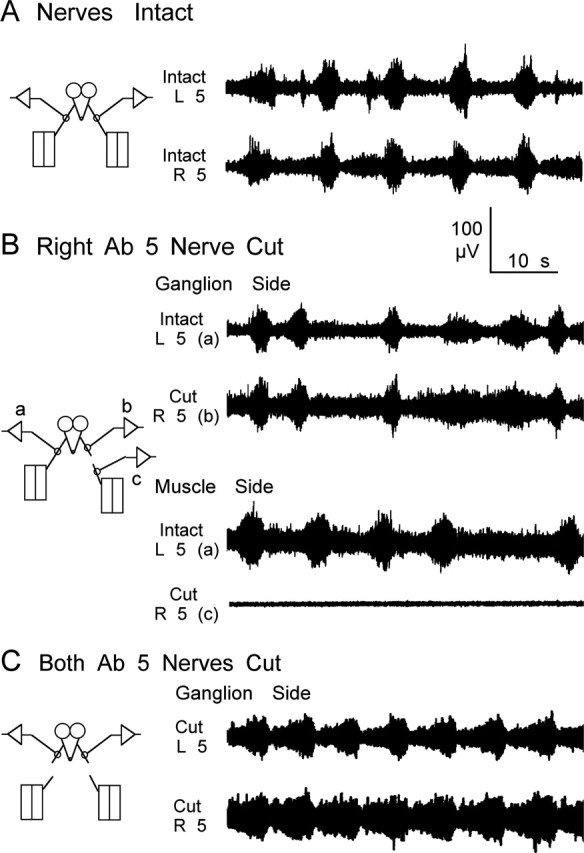
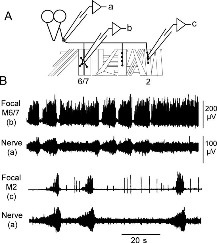
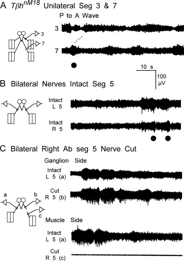
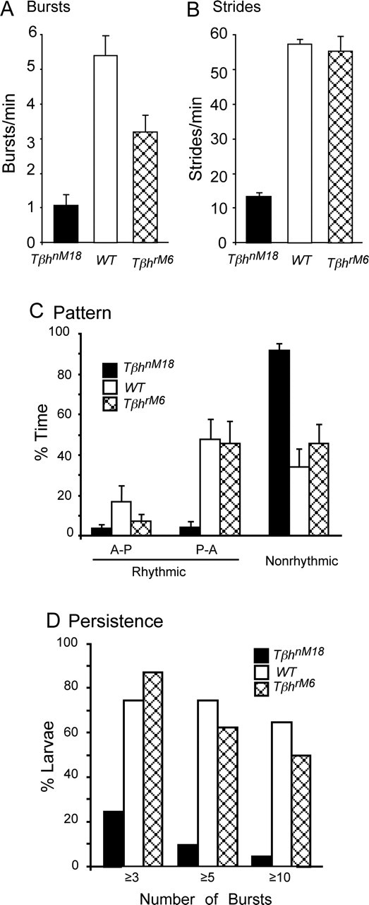
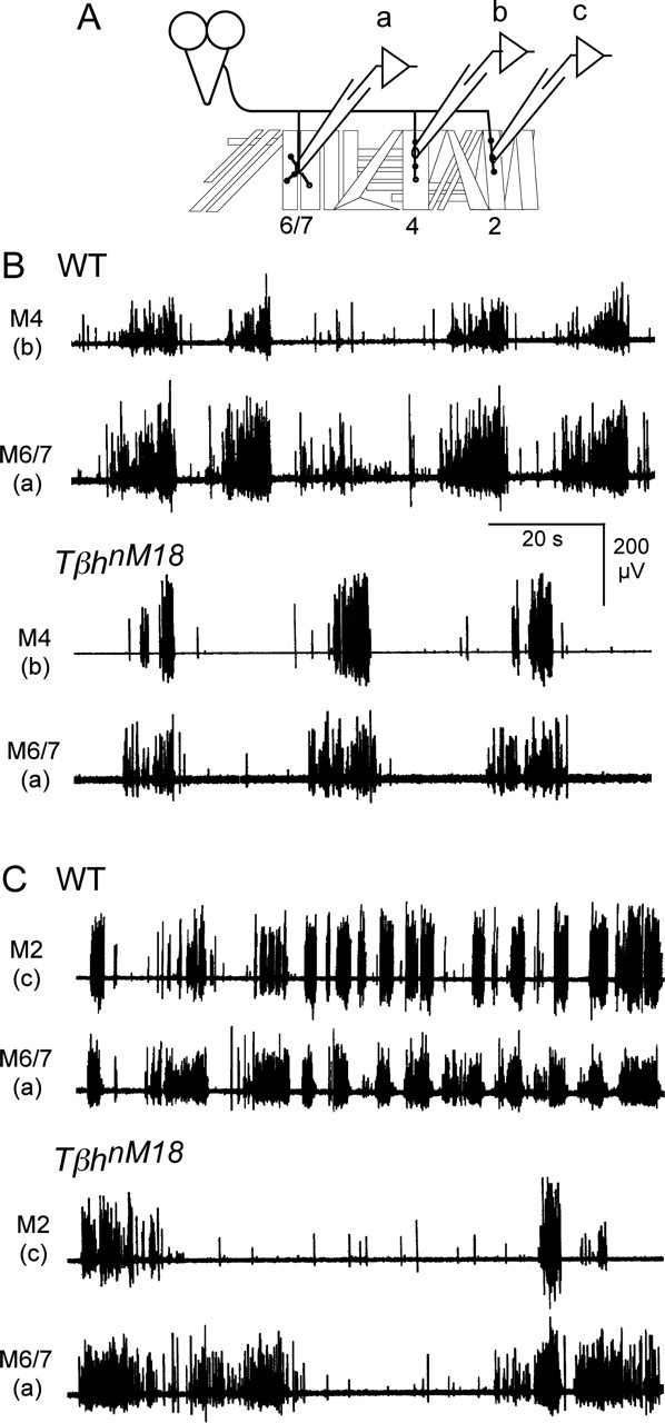
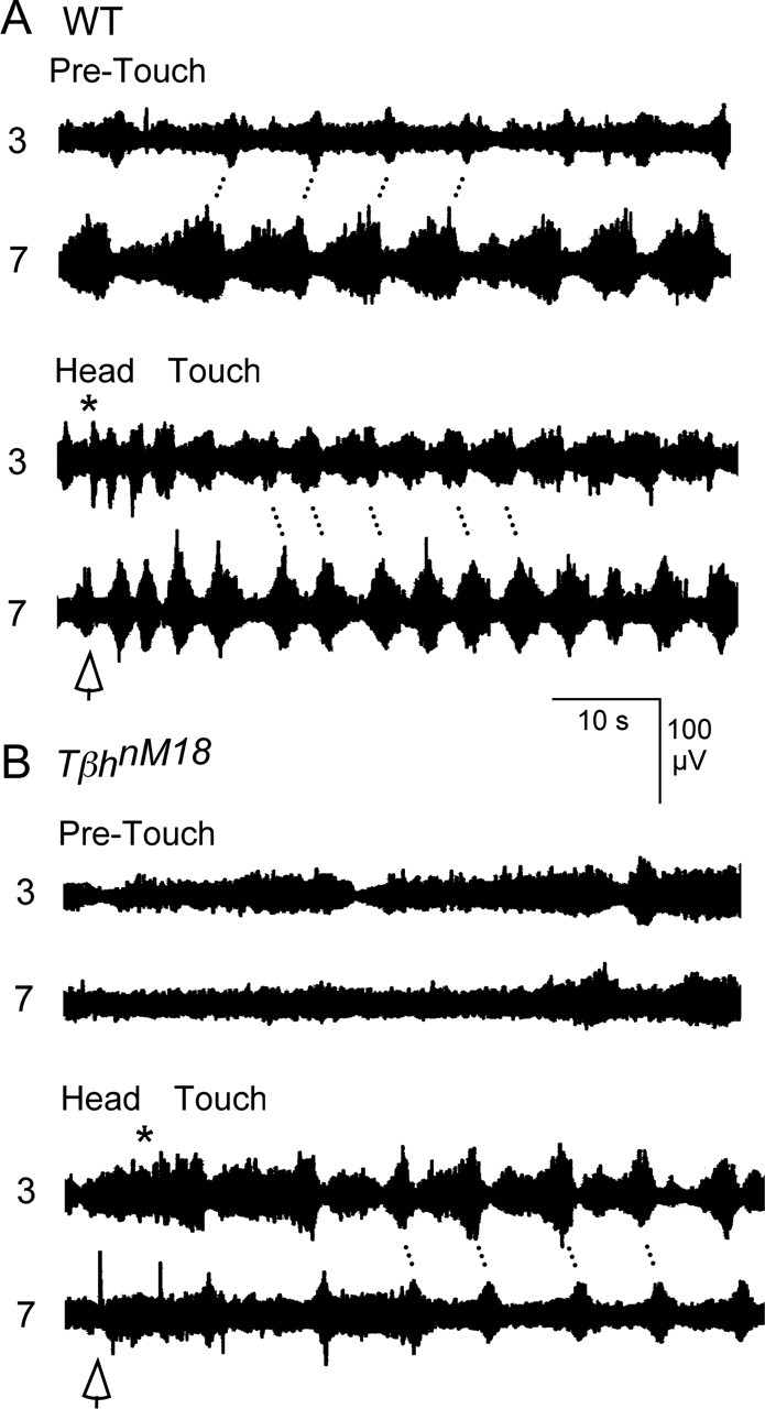
Forward locomotion of Drosophila melanogaster larvae is composed of rhythmic waves of contractions that are thought to be produced by segmentally organized central pattern generators. We present a systematic description of spike activity patterns during locomotive contraction waves in semi-intact wild-type and mutant larval preparations. We have shown previously that Tbetah(nM18) mutants, with altered levels of octopamine and tyramine, have a locomotion deficit. By recording en passant from the segmental nerves, we investigated the coordination of the neuronal activity driving contraction waves of the abdominal body-wall muscles. Rhythmic bursts of activity that occurred concurrently with locomotive waves were frequently observed in wild-type larvae but were rarely seen in Tbetah(nM18) mutants. These centrally generated patterned activities were eliminated in the distal stumps of both wild-type and Tbetah(nM18) larvae after severing the segmental nerve from the CNS. Patterned activities persisted in the proximal stumps deprived of sensory feedback from the periphery. Simultaneous recordings demonstrated a delay in the bursting activity between different segments, with greater delay for segments that were farther apart. In contrast, bilateral recordings within a single segment revealed a well synchronized activity pattern in nerves innervating each hemisegment in both wild-type and Tbetah(nM18) larvae. Significantly, rhythmic patterns of bursts and waves could be evoked in Tbetah(nM18) mutants by head or tail stimulation despite their highly irregular spontaneous activities. These observations suggest a role of the biogenic amines in the initiation and modulation of motor pattern generation. The technique presented here can be readily extended to examine the locomotion motor program of other mutants.


Figures

Figure 1.
Figure 1.
Representative crawling patterns and segmental nerve activity of WT, TβhrM6, and TβhnM18 Drosophila larvae. Larval position and outlines were captured by video recording for a duration of 2 min, digitized at 2 fps, and depicted by perimeter stacks. TβhnM18 mutant larvae traveled less distance than WT or TβhrM6 revertant larvae. The distance traveled by WT and TβhrM6 larvae were similar. The beginning of locomotion is marked by an asterisk. En passant recordings made from the nerve innervating abdominal segment 3 from semi-intact preparations of larvae in the wandering stage are shown. Activity recorded from WT and TβhrM6 nerves was organized into regular rhythmic bursts, whereas recordings from TβhnM18 mutants showed a high level of tonic nerve activity with very few bursts.
Figure 2.
Figure 2.
Correlation of nerve activity with waves of muscle contractions in WT larvae. A, Diagram of Drosophila larval neuromuscular preparation. Larval brain hemispheres (B), ventral ganglia (VG), pharyngeal muscles (PM), mouth hook (MH), and innervation of segmental body-wall muscles are indicated. Note the stereotypic pattern of the body-wall muscles in abdominal segments 2–7. B, Photograph of semi-intact Drosophila larval neuromuscular preparation showing en passant suction electrode placement. A recording from abdominal segmental nerves 3 and 7 is as indicated. C, D, Dissected larvae with the CNS intact produced regular rhythmic waves of muscle contractions during simultaneous video recordings and electrophysiological recordings. C, Selected frames from a video recording are in the top panel, and waves are diagramed in the bottom panel. In the initial phase of the contraction cycle, all of the body-wall muscles were relaxed (1), then the posterior segments were contracted (2). The contraction proceeded toward the head in a wave with medial (3) and anterior (4) segments contracting. Finally, all of the muscles relaxed before the next wave (5). D, Contractions were preceded by an increase in neuronal activity recorded from the segmental nerves that innervate the segment. Bilateral recordings were from left abdominal hemisegment 3 (L 3) and right hemisegment 7 (R 7). The time reference to contraction phases (1–5) during the cycle is indicated.
Figure 3.
Figure 3.
Bilateral and intersegmental coordination of neuronal activity recorded from segmental nerves of WT larvae. A, En passant recordings made bilaterally from segmental nerves innervating segment 5. Synchronized bursting activity was observed from the right (R) and left (L) sides associated with waves of contractions. B, En passant recordings made unilaterally from nerves innervating anterior segment 3 and posterior segment 7. There was a phase delay in activity recorded from nerves innervating anterior and posterior segments for unilateral recordings. Three common patterns of activity were observed in unilateral recordings: rhythmic activity recorded during anterior to posterior propagating contraction waves, rhythmic activity recorded during posterior to anterior waves, and nonrhythmic bursts unassociated with regular contraction waves. Recordings were from a single larva that spontaneously switched between the patterns. Note that the bursts tend to be longer in the body region where the wave originates. Muscle contraction waves here and in the following figures are marked with black dots. Seg, Segment.
Figure 4.
Figure 4.
Delay between bursts recorded from anterior and posterior segmental nerves. A, Nerve activity was recorded bilaterally during regular posterior to anterior waves of contractions, and the electrode was moved to other more posterior segments within a single larva. The peak activity was simultaneous in the bilateral segmental nerve 3 recordings (top). Activity recorded from segmental nerve 5 preceded the activity from segmental nerve 3 (middle), and the interval was increased further in recordings from segmental nerve 7 (bottom). B, Segmental delay during an anterior to posterior locomotive wave. Activity recorded from segmental nerve 3 preceded the activity from segmental nerve 7. L, Left; R, right.
Figure 5.
Figure 5.
Central origins of rhythmic activity recorded from segmental nerves of WT larvae. A, Activity recorded bilaterally from segment 5 with both nerves intact was coordinated and in phase. B, Cutting the nerve had little effect on the bursting activity recorded from the severed nerve attached to the ventral ganglion, whereas activity recorded from the nerve connected to the body wall ceased. C, Bursting activity was still coordinated and in phase even after bilaterally cutting the nerve to segment 5. A and B were from the same larva, and C is a representative example from a different larva. L, Left; R, right; Ab, abdominal.
Figure 6.
Figure 6.
A role of peripheral input in generating the regular rhythmic motor patterns in WT larvae. A, Unilateral recordings from anterior segmental nerve 3 and posterior nerve 6 revealed that cutting the nerves to all of the abdominal segments on the contralateral side had little effect on the bursting pattern. B, Activity was still organized into bursts when all of the abdominal nerves were cut, but the bursts were not as regular. C, D, Reducing peripheral input further by separating the brain from the body wall did not eliminate the burst-like activity. Unilateral recordings were made from anterior segmental nerve 3 and posterior nerve 6. Preparations consisting of the brain, pharyngeal muscles, and mouth hooks (C) or the isolated brain alone (D) had periods in which the activity was organized into bursts and showed an increase in tonic activity. L, Left; R, right; Ab, abdominal.
Figure 7.
Figure 7.
Synaptic activity recorded focally from motor terminals regions on muscles corresponds to activity recorded from nerves. A, Diagram of body-wall muscles within an abdominal hemisegment showing focal electrode placement. B, Simultaneous en passant recordings from segmental nerves and focal recordings from the nerve entry points of dorsal (M2) or ventral (M6/7) muscles indicated that nerve and muscle activity was coordinated and in phase. Note that the activity recorded from an individual muscle accounts for only part of the activity recorded from the nerve.
Figure 8.
Figure 8.
Disruption of locomotor rhythm in TβhnM18 mutants with a drastic decrease in rhythmic bursting activity and waves of contractions. A, Activity recorded unilaterally from two segmental nerves. There was a phase delay in activity recorded from anterior nerve 3 and posterior nerve 7. Note the increased tonic firing, fewer bursts, and fewer waves. B, Activity recorded bilaterally from segment 5 was coordinated and in phase. C, Cutting the nerve had little effect on the bursting activity recorded from the proximal stump attached to the ventral ganglion but eliminated activity in the distal stump connected to the body wall, indicating that central neurons produce patterned activity recorded from segmental nerves of TβhnM18 mutants. L, Left; R, right; Seg, segment; Ab, abdominal.
Figure 9.
Figure 9.
Burst frequency and the persistence of bursting episodes are reduced in TβhnM18 mutants. A, Summary of the number of bursts produced by WT, TβhrM6 revertants, and TβhnM18 larvae. All of the bursts associated with waves were counted regardless of the direction of propagation from TβhnM18 mutants (20 larvae, ∼200 min), WT larvae (20 larvae, ∼200 min), and TβhrM6 revertants (9 larvae, ∼50 min). TβhnM18 mutants had 80% fewer bursts per minute than the WT larvae and 65% fewer than TβhrM6 revertants (p < 0.001; Student’s t test). B, Summary of the number of strides produced by intact WT and TβhnM18 larvae crawling on agarose plates. TβhnM18 mutants had ∼80% fewer strides per minute than both the WT larvae and TβhrM6 revertants (WT, n = 47; TβhnM18, n = 79; TβhrM6, n = 19). Stride data are from Saraswati et al. (2004). C, Summary of time spent in anterior to posterior waves (A-P), posterior to anterior waves (P-A), or nonrhythmic bursting unassociated with rhythmic propagating contractions. TβhnM18 mutants spent significantly more time producing nonrhythmic contractions than WT larvae or TβhrM6 revertants (p < 0.001). Note that WT larvae (n = 20) and TβhrM6 revertants (n = 9) generated more P-A waves than A-P waves (p < 0.01), whereas time was evenly distributes between A-P and P-A waves for TβhnM18 mutants (p > 0.46, n = 20). There was no statistical difference for WT larvae or TβhrM6 revertants in the time spent among the patterns. Data were compiled for a 3 min period from each larva. D, Summary of the number of episodes that consisted of at least 3, 5, and 10 bursting events associated with contraction waves produced by WT, TβhrM6 and TβhnM18 larvae. TβhnM18 mutants (n = 20) produced significantly less bursts per episode than WT (n = 20) and TβhrM6 (n = 8) larvae (WT: p < 0.001 for 3, 5, and 10 bursts; TβhrM6: p < 0.001 for 3 bursts, p < 0.01 for 5 bursts, p < 0.05 for 10 bursts; t test). Data plotted in A–C are average ± SEM.
Figure 10.
Figure 10.
Correlation of activity patterns between dorsal, lateral, and ventral muscles within a hemisegment of WT larvae and TβhnM18 mutants. A, Diagram of body-wall muscles within an abdominal hemisegment showing focal electrode placement. B, Simultaneous focal recording from muscles within the same hemisegment indicated that ventral muscles 6 and 7 (M6/7) and lateral muscle 4 (M4) tend to be activated simultaneously. C, Ventral (M6/7) and dorsal muscle 2 (M2) muscles also have similar activity patterns in both WT larvae and TβhnM18 mutants. Despite the concurrent activity observed in each muscle pair, the pattern was similar but not identical within each pair, suggesting that they are innervated by different neurons. Note the weaker correlation of activity between different muscles of TβhnM18 mutant larvae.
Figure 11.
Figure 11.
A, B, Motor patterns evoked by tactile stimulation in WT larvae (A) and TβhnM18 mutants (B). The anterior end of the larval preparations was lightly tapped with a silver wire probe. Before touching, the ongoing nerve activity consisted of bursts originating in segment 7 that were followed by bursts in segment 3 in WT larvae and nonrhythmic bursting in TβhnM18 mutants (pre-touch). Touching the head stopped the ongoing activities (open arrows) and induced a burst observed in segmental nerve 3 that was not associated with a wave (asterisk). The relative timing of the nerve activity reversed for WT larvae, indicating a conversion of posterior to anterior contraction waves to anterior to posterior waves. Significantly, tactile stimulation evoked a series of persistent alternating bursts in segment 3 and 7 of TβhnM18 mutants, associated with anterior to posterior waves that were rarely encountered in the spontaneous activity of the mutant larvae.

Similar articles

Cited by

References

    1. Alkema MJ, Hunter-Ensor M, Ringstad N, Horvitz HR (2005). Tyramine functions independently of octopamine in the Caenorhabditis elegans nervous system. Neuron 46:247–260. - PubMed
    1. Baier A, Wittek B, Brembs B (2002). Drosophila as a new model organism for the neurobiology of aggression? J Exp Biol 205:1233–1240. - PubMed
    1. Barclay JW, Atwood HL, Robertson RM (2002). Impairment of central pattern generation in Drosophila cysteine string protein mutants. J Comp Physiol A Neuroethol Sens Neural Behav Physiol 188:71–78. - PubMed
    1. Belanger JH, Trimmer BA (2000). Combined kinematic and electromyographic analyses of proleg function during crawling by the caterpillar Manduca sexta. J Comp Physiol A Neuroethol Sens Neural Behav Physiol 186:1031–1039. - PubMed
    1. Berrigan D, Pepin DJ (1995). How maggots move: allometry and kinematics of crawling in larval diptera. J Insect Physiol 41:329–337.

Publication types

MeSH terms

LinkOut - more resources