. 2006 Feb 1;26(5):1551-61.
doi: 10.1523/JNEUROSCI.3101-05.2006.

The endocannabinoid system promotes astroglial differentiation by acting on neural progenitor cells

Affiliations

The endocannabinoid system promotes astroglial differentiation by acting on neural progenitor cells

Tania Aguado et al. J Neurosci. .

Abstract

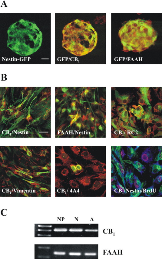
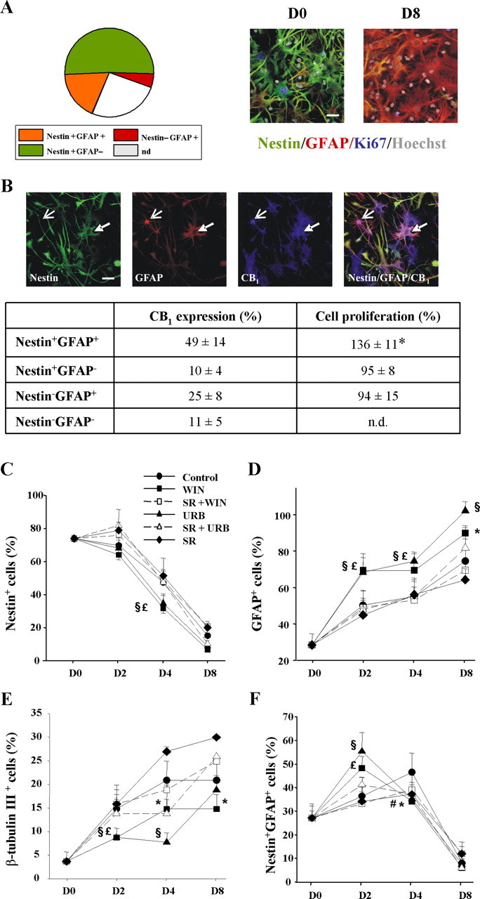
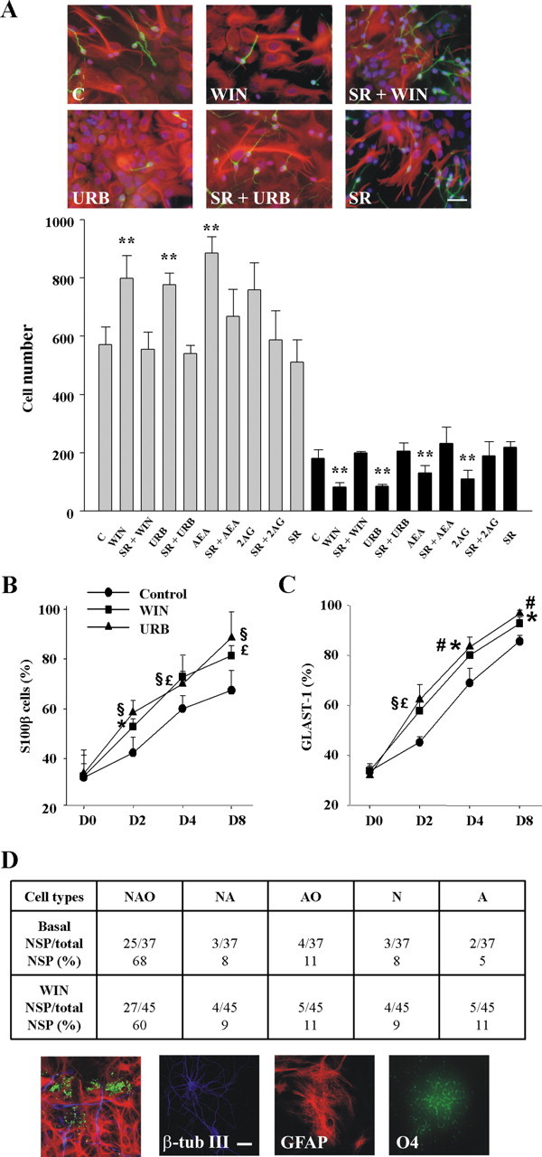
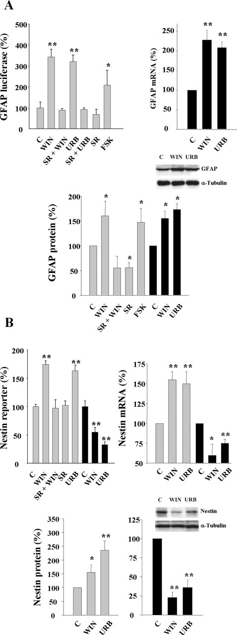
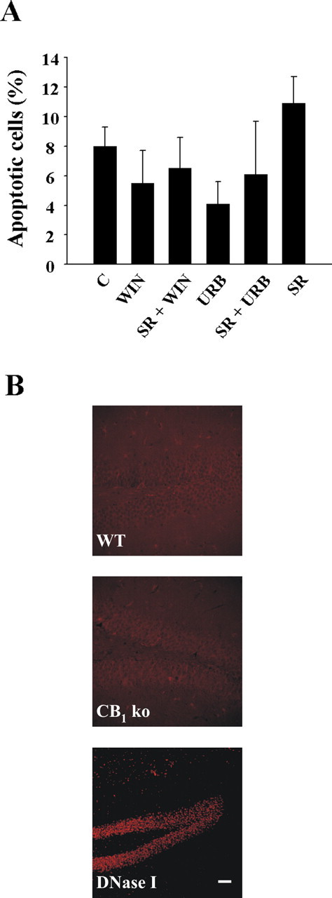
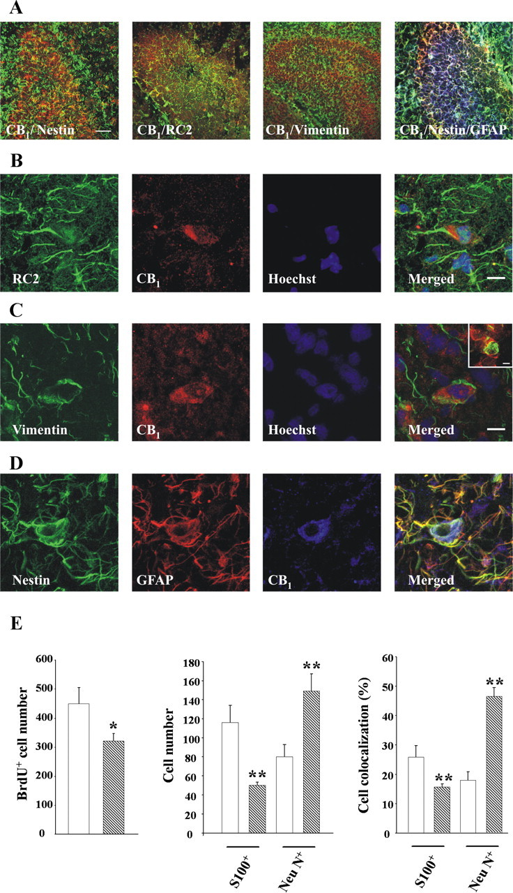
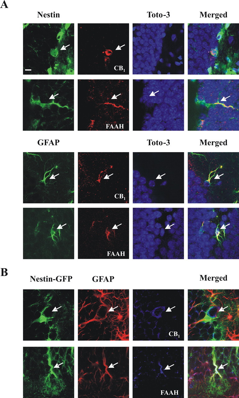
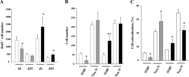
Endocannabinoids exert an important neuromodulatory role via presynaptic cannabinoid CB1 receptors and may also participate in the control of neural cell death and survival. The function of the endocannabinoid system has been extensively studied in differentiated neurons, but its potential role in neural progenitor cells remains to be elucidated. Here we show that the CB1 receptor and the endocannabinoid-inactivating enzyme fatty acid amide hydrolase are expressed, both in vitro and in vivo, in postnatal radial glia (RC2+ cells) and in adult nestin type I (nestin(+)GFAP+) neural progenitor cells. Cell culture experiments show that CB1 receptor activation increases progenitor proliferation and differentiation into astroglial cells in vitro. In vivo analysis evidences that, in postnatal CB1(-/-) mouse brain, progenitor proliferation and astrogliogenesis are impaired. Likewise, in adult CB1-deficient mice, neural progenitor proliferation is decreased but is increased in fatty acid amide hydrolase-deficient mice. In addition, endocannabinoid signaling controls neural progenitor differentiation in the adult brain by promoting astroglial differentiation of newly born cells. These results show a novel physiological role of endocannabinoids, which constitute a new family of signaling cues involved in the regulation of neural progenitor cell function.


Figures

Figure 1.
Figure 1.
The endocannabinoid system is expressed in postnatal neural progenitors. A, Immunofluorescence was performed to assess CB1 and FAAH (red) expression in nestin-GFP-derived neurospheres. Scale bar, 20 μm. B, Immunofluorescence was performed with CB1 or FAAH antibodies (red) in combination with nestin, RC2, vimentin, or phosphorylated vimentin (4A4) antibodies (green). In addition, triple labeling was performed with CB1 (blue), nestin (green), and BrdU (red) antibodies. Scale bars, 20 μm. C, The presence of CB1 receptor and FAAH mRNA was determined by RT-PCR in NPs, differentiated neurons (N), and differentiated astroglial cells (A).
Figure 2.
Figure 2.
Characterization of postnatal progenitor proliferation and astroglial differentiation. A, Distribution of the phenotypic analysis with the indicated cell markers of proliferating (Ki67+) cells after 16 h of differentiation. Representative immunofluorescence pictures of NPs at different stages of differentiation using the indicated markers (n.d., not determined). Scale bar, 30 μm. B, CB1 receptor distribution in the different cellular phenotypes (as above). Representative nestin+GFAP+ CB1+ (filled arrow) and nestinGFAP+ CB1+ cells (open arrow) are shown. Scale bar, 30 μm. Quantification of CB1 distribution compared with the proliferative effect induced by WIN-55,212-2 for each cell type. Significantly different from controls, *p < 0.01. C–F, Cannabinoid impact on NP differentiation. Quantification of the percentage of nestin+ (C), GFAP+ (D), β-tubulin III+ (E), and nestin+GFAP+ (F) cells versus total cell number at the indicated differentiation times in the presence of vehicle, WIN-55,212-2, or URB597 alone or with SR141716, and SR141716 alone. Results correspond to three independent experiments. Significant differences between cannabinoid-treated versus control cells are shown. *WIN-55,212-2, #URB597, p < 0.05; £WIN-55,212-2, §URB597, p < 0.01.
Figure 3.
Figure 3.
Cannabinoids promote astroglial differentiation in vitro. A, Immunofluorescence with anti-β-tubulin III (green) and GFAP (red) antibodies after a 48 h differentiation period of NP cells in the presence of vehicle (C), 30 nm WIN-55,212-2 alone or with 2 μm SR141716, 30 nm URB597 alone or with SR141716, and SR141716 alone. Scale bar, 30 μm. Quantification of the GFAP+ (gray bars) and β-tubulin III+ (black bars) after differentiation of NPs as above or in the presence of 10 μm AEA or 2AG. Results correspond to three independent experiments. *p < 0.05; **p < 0.01. Quantification of the percentage of S100β+ (C) or GLAST-1+ (D) cells versus total cell number at the indicated times in the presence of vehicle, WIN-55,212-2, or URB597. *WIN-55,212-2, #URB597, p < 0.05; £WIN-55,212-2, §URB597, p < 0.01. D, Clonal analysis of postnatal NP-derived neurospheres indicating the number of clones that generated neurons (N), astrocytes (A), and oligodendrocytes (O) together with other lineage-restricted clones. Triple immunostaining with β-tubulin III, GFAP, and O4 antibodies was performed after 8 d differentiation. Examples of differentiated cells are shown. Scale bar, 20 μm.
Figure 4.
Figure 4.
Cannabinoid regulation of GFAP and nestin expression. A, Luciferase activity under the control of the GFAP promoter (left) was determined in transfected NPs after 24 h incubation in the presence of the indicated stimuli. Forskolin (FSK; 5 μm) was used as a positive control. GFAP mRNA levels (right) were determined after cannabinoid stimulation for 4 d. GFAP protein expression (bottom) with the indicated stimuli was determined after 24 h (gray bars) or 4 d (black bars). B, Nestin reporter activity (left) was determined in transfected NPs after 24 h (gray bars) or 4 d (black bars) as above. Nestin mRNA levels (right) and protein levels (bottom) after cannabinoid stimulation for 24 h (gray bars) or 4 d (black bars). Densitometric quantification is referred to the corresponding α-tubulin signal. Results correspond to three independent experiments. Representative Western blots are shown. Significantly different from controls, *p < 0.05; **p < 0.01.
Figure 5.
Figure 5.
Lack of cannabinoid-induced neural progenitor cell death. A, Quantification of TUNEL staining in NPs stimulated for 48 h with the indicated cannabinoid agonists or antagonists (as in differentiation experiments). B, Apoptosis was determined by TUNEL staining in adult hippocampal brain sections in CB1 knock-out mice (KO) and their wild-type littermates (WT) (n = 5 for each group; representative pictures are shown). DNase-treated sections were used as positive control. Scale bar, 35 μm.
Figure 6.
Figure 6.
Role of the endocannabinoid system in postnatal astrogliogenesis and neurogenesis in vivo. A, Low-magnification images of CB1 expression in postnatal hippocampus sections stained for nestin, RC2, and vimentin, or nestin and GFAP. Scale bar, 40 μm. B–D, CB1 expression (red) in progenitor cells as identified in green color by RC2 (B) and vimentin (C) immunoreactivity. Double-labeled nestin+ (green) GFAP+ (red) cells (D) also express the CB1 receptor (blue). Cells were counterstained with Hoechst (blue). Scale bar, 10 μm. Inset shows coexpression of CB1 and phosphorylated vimentin. Scale bar, 6 μm. E, Number of BrdU+ cells per section (left), number of BrdU+ cells that colocalize with S100β or NeuN (middle), and percentage of BrdU+ cells that coexpress S100β or NeuN (right) in the hippocampus of CB1 knock-out mice (hatched bars) and their wild-type littermates (white bars). Significantly different from controls, *p < 0.05; **p < 0.01.
Figure 7.
Figure 7.
Expression of the CB1 receptor and FAAH in adult hippocampal NPs in vivo. A, Expression of CB1 and FAAH (red) assessed by confocal microscopy as revealed by nestin or GFAP immunoreactivity (green) in mouse hippocampal sections. Cells were counterstained with TOTO-3 iodide (blue). Scale bar, 8 μm. B, Triple labeling showing the expression of CB1 and FAAH (blue) in GFAP-immunoreactive (red) nestin-GFP+ cells. Representative images are shown.
Figure 8.
Figure 8.
Role of the endocannabinoid system in adult astrogliogenesis and neurogenesis in vivo. A, Number of BrdU+ cells per mouse in the hippocampus of CB1 knock-out mice (hatched bars), FAAH knock-out mice (black bars), and their respective wild-type littermates (white bars) injected with BrdU and perfused 1 or 25 d later. B, Number of BrdU+ cells that colocalize with S100β or NeuN 25 d after the BrdU pulse. C, Percentage of BrdU+ cells that coexpress S100β or NeuN. Significantly different from controls, *p < 0.05; **p < 0.01.

References

    1. Aguado T, Monory K, Palazuelos J, Stella N, Cravatt B, Lutz B, Marsicano G, Kokaia Z, Guzmán M, Galve-Roperh I (2005). The endocannabinoid system drives neural progenitor proliferation. FASEB J 19:1704–1706. - PubMed
    1. Alvarez-Buylla A, Lim D (2004). For the long run: maintaining germinal niches in the adult brain. Neuron 41:683–686. - PubMed
    1. Cravatt BF, Demarest K, Patricelli MP, Bracey MH, Giang DK, Martin BR, Lichtman AH (2001). Supersensitivity to anandamide and enhanced endogenous cannabinoid signaling in mice lacking fatty acid amide hydrolase. Proc Natl Acad Sci USA 98:9371–9376. - PMC - PubMed
    1. Doetsch F, Caille I, Lim DA, Garcia-Verdugo JM, Alvarez-Buylla A (1999). Subventricular zone astrocytes are neural stem cells in the adult mammalian brain. Cell 97:703–716. - PubMed
    1. Egertova M, Cravatt BF, Elphick MR (2003). Comparative analysis of fatty acid amide hydrolase and cb(1) cannabinoid receptor expression in the mouse brain: evidence of a widespread role for fatty acid amide hydrolase in regulation of endocannabinoid signaling. Neuroscience 119:481–496. - PubMed

Publication types

MeSH terms

Substances