. 2006 Feb 1;26(5):1588-95.
doi: 10.1523/JNEUROSCI.3979-05.2006.

Simultaneous release of glutamate and acetylcholine from single magnocellular "cholinergic" basal forebrain neurons

Affiliations

Simultaneous release of glutamate and acetylcholine from single magnocellular "cholinergic" basal forebrain neurons

Timothy G J Allen et al. J Neurosci. .

Abstract

Basal forebrain (BF) neurons provide the principal cholinergic drive to the hippocampus and cortex. Their degeneration is associated with the cognitive defects of Alzheimer's disease. Immunohistochemical studies suggest that some of these neurons contain glutamate, so might also release it. To test this, we made microisland cultures of single BF neurons from 12- to 14-d-old rats. Over 1-8 weeks in culture, neuronal processes made autaptic connections onto the neuron. In 34 of 36 cells tested, a somatically generated action potential was followed by a short-latency EPSC that was blocked by 1 mM kynurenic acid, showing that they released glutamate. To test whether the same neuron also released acetylcholine, we placed a voltage-clamped rat myoball expressing nicotinic receptors in contact with a neurite. In six of six neurons tested, the glutamatergic EPSC was accompanied by a nicotinic (hexamethonium-sensitive) myoball current. Stimulation of the M2-muscarinic presynaptic receptors (characterized using tripitramine and pirenzepine) produced a parallel inhibition of autaptic glutamatergic and myoball nicotinic responses; metabotropic glutamate receptor stimulation produced similar but less consistent and weaker effects. Atropine enhanced the glutamatergic EPSCs during repetitive stimulation by 25 +/- 6%; the anti-cholinesterase neostigmine reduced the train EPSCs by 37 +/- 6%. Hence, synaptically released acetylcholine exerted a negative-feedback inhibition of coreleased glutamate. We conclude that most cholinergic basal forebrain neurons are capable of releasing glutamate as a cotransmitter and that the release of both transmitters is subject to simultaneous feedback inhibition by synaptically released acetylcholine. This has implications for BF neuron function and for the use of cholinesterase inhibitors in Alzheimer's disease.


Figures

Figure 1.
Figure 1.
A, A typical microisland visualized using Normarski differential interference contrast optics. The neuron shown was obtained from a 12-d-old rat and had been maintained in culture for 22 d. Scale bar, 50 μm. B, Somatic action potentials generated by a brief (10 ms, 400 pA) current injection are followed by autaptic EPSPs, one of which generates a second action potential. The autaptic EPSP is attenuated by 1 mm kynurenic acid. [The residual, kynurenic acid-insensitive ADP results from activation of calcium-activated chloride channels (Sim and Allen 1998).] C, The EPSPs were prolonged when NMDA-mediated responses were enhanced by removal of extracellular Mg2+ and the addition of 3 μm glycine. (Note the slower time base.) ctrl, Control. D, Voltage-clamp recording of autaptic EPSCs in 0 [Mg2+], 1 μm glycine solution. The EPSC comprises fast and slow components. The NMDA antagonist d-AP-5 (100 μm) selectively inhibited the slow component; the subtracted d-AP-5-sensitive current shows the characteristic slow rise time and decay of an NMDA current. The residual fast component was inhibited by the AMPA receptor antagonist NBQX (100 μm).
Figure 2.
Figure 2.
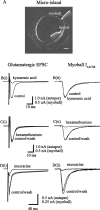
Corelease of glutamate and acetylcholine from single magnocellular basal forebrain neurons grown in microisland culture and suppression of their release by stimulating mAChRs. A, Photographic montage of a single-cell microisland from which the simultaneous release of glutamate (detected in the form of an autaptic EPSC by the neuronal recording electrode) and ACh (detected in the form of a nicotinic current in a voltage-clamped skeletal myoball placed in contact with the neurites of the cell) were detected from the same cell. Scale bar, 30 μm. Bi, Ci, Di, Glutamatergic autaptic EPSCs evoked by single action potentials elicited in response to a brief (2 ms) depolarizing step from a holding potential of −70 mV. Bii, Cii, Dii, Simultaneous release of ACh from the same cells detected using single perforated-patched skeletal myoballs voltage clamped at −70 mV and placed in contact with the neurites of the cell (Allen, 1999). Note that the records shown in B and C were obtained from the same neuron/myoball pair; records in D are from a different neuron/myoball pair. In B, the glutamate antagonist kynurenic acid (1 mm) abolished the autaptic EPSC without affecting the myoball response. Conversely, in C, the nicotinic ACh-receptor antagonist hexamethonium (100 μm) abolished the response of the myoball but had almost no effect on the autaptic EPSC. In D, the mAChR agonist muscarine (10 μm) reversibly inhibits both glutamate and ACh release from a single neuron.
Figure 3.
Figure 3.
Muscarinic receptor-mediated inhibition of the glutamatergic autaptic EPSC. A, EPSCs in a single cultured neuron in response to single action potentials elicited by imposing brief (2 ms) depolarization steps from Vh −80 mV under control conditions and in the presence of increasing concentrations of muscarine (0.1–10 μm). Each response is an average of four to six EPSCs after equilibration at each of the different agonist concentrations. B, Dose–response relationships for inhibition of EPSC by ACh recorded under control conditions and the presence of the M1/M4 mAChR antagonist pirenzepine or the M2 antagonist tripitramine. Inhibition was measured as percentage reduction of peak EPSC amplitude. Curves are least-squares fits to the following Hill equation: y = ymax × xnH/ (xnH + KnH), where y = percentage inhibition, ymax = 100, x = agonist concentration (conc; micromolar), K is a constant (equivalent to the IC50; micromolar), and nH is the Hill slope. Fitted curves were constructed using the mean IC50 and Hill slope values obtained from the individual cells. All data points are the mean ± SEM values obtained after normalization of data obtained from the individual cells (n = 5–7 cells for each point). Mean values for K (micromolar; nH in parentheses) were as follows: control, 0.395 ± 0.152 (slope, 0.95); pirenzepine, 0.639 ± 0.017 (slope, 1.23); and tripitramine, 8.47 ± 2.4 (slope, 0.93).
Figure 4.
Figure 4.
Glutamate-mediated EPSCs are enhanced when muscarinic autoreceptors are blocked by atropine, showing that glutamate release is depressed by coreleased ACh. A, B, Autaptic glutamatergic EPSCs evoked at 2 Hz under control conditions and in the presence of the mAChR antagonist atropine (30 nm). C, Mean amplitudes of the second to 20th EPSCs in A and B recorded before and after adding atropine. D, A brief burst of four glutamatergic EPSCs evoked at a frequency of 10 Hz followed by a delay of 1 s before a final (5th) EPSC, recorded before and after adding 100 nm atropine. (This protocol was repeated every 60 s.) E, F, The initial four (*) and final (5th; **) EPSCs on an expanded scale. In the presence of atropine, the amplitudes of the first and second EPSCs remained unchanged, whereas the amplitude of the subsequent EPSCs became larger. This shows that the presynaptic muscarinic autoreceptors activate within 150–200 ms of the start of the first action potential/release event. Once activated in this way, functional inhibition of glutamate release persists for a period in excess of 1 s (F). EPSCs were evoked by briefly stepping to a depolarized potential for 2 ms from a holding potential of −70 mV; all traces are the mean of three or four repetitions. Note that the dramatic decline in the glutamatergic EPSC after the first stimulus mostly persisted in the presence of atropine and therefore could not be attributed to cholinergic presynaptic inhibition. One possibility is that it results from AMPA receptor desensitization. We tested this using 200 μm diazoxide (to reduce desensitization) (Yamada and Tang, 1993) in the presence of 100 μm tolbutamide (the latter to block KATP channel activation) using the protocol in D. Diazoxide increased both amplitude and half-decay times of the first EPSC [by 51 ± 6.4% (n = 9) and 212 ± 34.7% (n = 7), respectively] but did not significantly affect the depression of the second EPSC (second/first EPSC amplitudes 35 ± 5.5 and 43.4 ± 5.7% in the absence and presence of drug, respectively; n = 8). Because the release of ACh from these neuron processes shows a comparable fall-off (Allen and Brown 1996), it most likely results from a temporary depletion of transmitter or exhaustion of the release machinery. The 1 s rest period in D allows some recovery from this depletion.
Figure 5.
Figure 5.
Effects of the anti-cholinesterase neostigmine (3 μm) on presynaptic muscarinic receptor-mediated inhibition of glutamatergic EPSCs. A, Four EPSCs were evoked at a frequency of 10 Hz, with a delay of 1 s before a fifth EPSC was elicited (B). This protocol repeated every 60 s. Addition of neostigmine had no effect on the initial two EPSCs but depressed all subsequent ones, including that recorded 1 s later (B, C). Addition of atropine (100 nm) in the presence of neostigmine fully reversed this effect and even enhanced the later EPSCs.
Figure 6.
Figure 6.
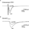
Stimulation of presynaptic metabotropic glutamate receptors (mGluRs reduces the release of both glutamate and ACh). Records show the depressant effect of the mGluR agonist t-ACPD (30 μm) on glutamatergic autaptic EPSCs (A) and myoball nicotinic ACh currents (B), respectively, recorded simultaneously on stimulating a single BF neuron in a microisland culture (see Fig. 2). Note that all records are the mean of four repetitions in the presence and absence of agonist. For clarity, washout responses have been omitted, but autaptic and myoball responses recovered to 97 and 103%, respectively, of the control (ctrl) values.

References

    1. Allen TGJ (1999). The role of N-, Q- and R-type Ca2+ channels in feedback inhibition of ACh release from rat basal forebrain neurons. J Physiol (Lond) 515:93–107. - PMC - PubMed
    1. Allen TGJ, Brown DA (1993). M2 muscarinic receptor-mediated inhibition of the Ca2+ current in rat magnocellular cholinergic basal forebrain neurones. J Physiol (Lond) 466:173–189. - PMC - PubMed
    1. Allen TGJ, Brown DA (1996). Detection and modulation of acetylcholine release from neurites of rat basal forebrain cells in culture. J Physiol (Lond) 492:453–466. - PMC - PubMed
    1. Allen TGJ, Brown DA (2004). Modulation of the excitability of cholinergic basal forebrain neurones by KATP channels. J Physiol (Lond) 554:353–370. - PMC - PubMed
    1. Beaulieu C, Somogyi P (1991). Enrichment of cholinergic synaptic terminals on GABAergic neurons and coexistence of immunoreactive GABA and choline acetyltransferase in the same synaptic terminals in the striate cortex of the cat. J Comp Neurol 304:666–680. - PubMed

Publication types