. 2006 Feb 2;439(7076):612-6.
doi: 10.1038/nature04487.

Structure of epsilon15 bacteriophage reveals genome organization and DNA packaging/injection apparatus

Affiliations

Structure of epsilon15 bacteriophage reveals genome organization and DNA packaging/injection apparatus

Wen Jiang et al. Nature. .

Abstract

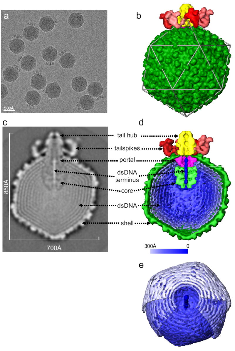
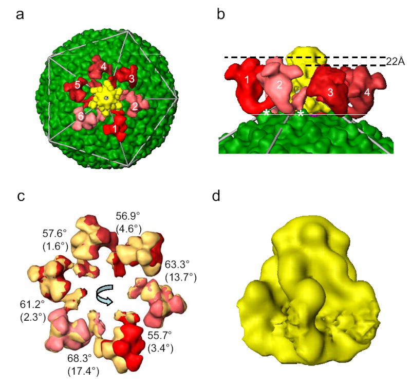
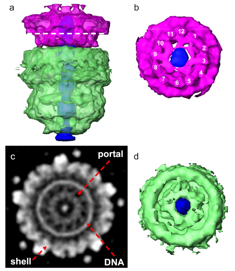
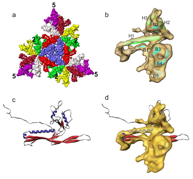
The critical viral components for packaging DNA, recognizing and binding to host cells, and injecting the condensed DNA into the host are organized at a single vertex of many icosahedral viruses. These component structures do not share icosahedral symmetry and cannot be resolved using a conventional icosahedral averaging method. Here we report the structure of the entire infectious Salmonella bacteriophage epsilon15 (ref. 1) determined from single-particle cryo-electron microscopy, without icosahedral averaging. This structure displays not only the icosahedral shell of 60 hexamers and 11 pentamers, but also the non-icosahedral components at one pentameric vertex. The densities at this vertex can be identified as the 12-subunit portal complex sandwiched between an internal cylindrical core and an external tail hub connecting to six projecting trimeric tailspikes. The viral genome is packed as coaxial coils in at least three outer layers with approximately 90 terminal nucleotides extending through the protein core and the portal complex and poised for injection. The shell protein from icosahedral reconstruction at higher resolution exhibits a similar fold to that of other double-stranded DNA viruses including herpesvirus, suggesting a common ancestor among these diverse viruses. The image reconstruction approach should be applicable to studying other biological nanomachines with components of mixed symmetries.


Figures

Figure 1
Figure 1. Structure of Epsilon15 phage
(a) 200 kV CCD image of Epsilon15 particles embedded in vitreous ice. (b) Surface rendering of the 20 Å resolution 3-D map of entire Epsilon15 phage reconstructed without symmetry imposition. The capsid (dark green) exhibits good icosahedral symmetry as indicated by the icosahedral lattice (gray). The structural components of Epsilon15 phage are annotated in the central section density (c) and the cut-away surface view (d) of the 3-D density map. Each of the structural components is colored differently and the same color scheme is used in Figures 1–3. (e) A slightly tilted view of the coaxially packed dsDNA genome. Only portions of the first and second layers of dsDNA were shown so that the three outermost layers can be viewed. Color gradient (light blue to darker blue) is used in (d) and (e) to indicate the likely packaging order of the dsDNA starting from outer layer towards inner layers and ending as the central straight segment of terminus.
Figure 2
Figure 2. The tail structure
(a) Top view of the tail in the Epsilon15 reconstruction. Each of the 6 tailspikes is uniquely colored with a random shade of red in order to illustrate the inexact 6-fold arrangement around the central tail hub (yellow) with good 6-fold symmetry. (b) Zoom-in side view of the tailspikes. The contact sites for two (labeled as 1 and 2) out of the six tailspikes are on the capsid surface protrusion densities at local 2-fold positions (labeled with *). (c) Each of the tailspikes is aligned to its neighbor (counter-clockwise) with indicated amount of rotation around an axis that is tilted away from the 6-fold axis in different degrees as indicated in parenthesis. The tailspikes in the original positions are displayed in the same shades of red colors as in (a) while the rotated tailspikes in the new positions are displayed in yellow. (d) Side view of the tail hub. The segmentation of the tail hub at the interacting regions with the tailspikes is arbitrary because of their tight binding and limited resolution.
Figure 3
Figure 3. Structure of portal complex and internal core
(a) Side view of the portal complex (pink) and internal core (light green) and the putative straight dsDNA terminus (dark blue). The portal complex and internal core are colored semi-transparently to show their central channels are filled with the putative dsDNA terminus. (b) Bottom view of the portal complex with the nearly 12-fold structural features labeled around the filled central channel. (c) The apparent 12-fold arrangement of densities for the portal complex in a section image of the 3-D map. The location of the section is indicated by the dashed line in (a). (d) Top view of the internal core (light green) showing central channel filled with the putative dsDNA terminus (dark blue).
Figure 4
Figure 4. Shell protein structure
(a) Surface rendering of three asymmetric units forming one icosahedral face of the T=7l shell. Each of the 7 subunits is colored differently. (b) A single averaged shell protein subunit with 3 helices (H1-H3) and 5 sheets (B1-B5) annotated. (c) Ribbon representation of the atomic model of HK97 head protein gp5 (PDB accession ID: 1OHG). (d) Superposition of Epsilon15 shell protein average subunit and the HK97 head protein gp5.

References

    1. McConnell M, Reznick A, Wright A. Studies on the initial interactions of bacteriophage epsilon15 with its host cell, Salmonella anatum. Virology. 1979;94:10–23. - PubMed
    1. Jiang W, et al. Coat protein fold and maturation transition of bacteriophage P22 seen at subnanometer resolutions. Nat Struct Biol. 2003;10:131–5. - PubMed
    1. Wikoff WR, et al. Topologically linked protein rings in the bacteriophage HK97 capsid. Science. 2000;289:2129–33. - PubMed
    1. Fokine A, et al. Structural and functional similarities between the capsid proteins of bacteriophages T4 and HK97 point to a common ancestry. Proc Natl Acad Sci U S A. 2005;102:7163–8. - PMC - PubMed
    1. Morais MC, et al. Conservation of the capsid structure in tailed dsDNA bacteriophages: the pseudoatomic structure of phi29. Mol Cell. 2005;18:149–59. - PubMed

Publication types