Review
. 2006 Feb;116(2):288-96.
doi: 10.1172/JCI27699.

Mechanisms of progression and regression of renal lesions of chronic nephropathies and diabetes

Affiliations
Review

Mechanisms of progression and regression of renal lesions of chronic nephropathies and diabetes

Giuseppe Remuzzi et al. J Clin Invest. 2006 Feb.

Abstract

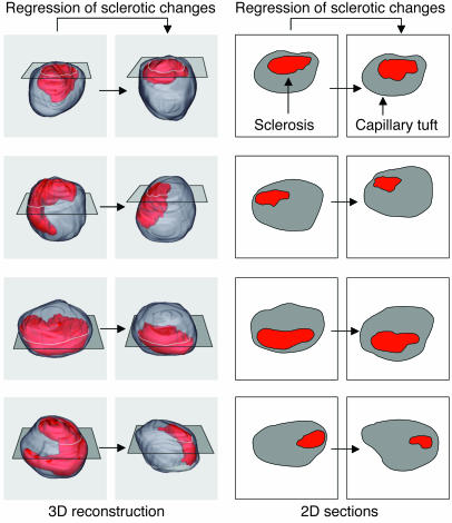
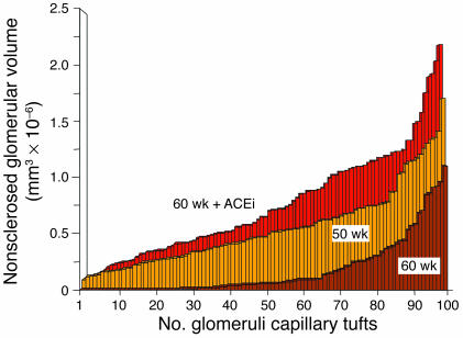
The incidence of chronic kidney diseases is increasing worldwide, and these conditions are emerging as a major public health problem. While genetic factors contribute to susceptibility and progression of renal disease, proteinuria has been claimed as an independent predictor of outcome. Reduction of urinary protein levels by various medications and a low-protein diet limits renal function decline in individuals with nondiabetic and diabetic nephropathies to the point that remission of the disease and regression of renal lesions have been observed in experimental animals and even in humans. In animal models, regression of glomerular structural changes is associated with remodeling of the glomerular architecture. Instrumental to this discovery were 3D reconstruction studies of the glomerular capillary tuft, which allowed the quantification of sclerosis volume reduction and capillary regeneration upon treatment. Regeneration of capillary segments might result from the contribution of resident cells, but progenitor cells of renal or extrarenal origin may also have a role. This review describes recent advances in our understanding of the mechanisms and mediators underlying renal tissue repair ultimately responsible for regression of renal injury.


Figures

Figure 1
Figure 1
The progressive nature of chronic kidney disease. The progression to ESRD, as underlined by the progressive decline of GFR, is highly variable. Here is reported the natural history of autosomal-dominant polycystic kidney disease (ADPKD) in patients with PKD1 mutation in the PKD1 gene as an example of genetic renal disease. Progressive renal disease occurs in 20–40% of patients with type 2 diabetes. Progression to renal failure occurs in 30% of patients with IgA nephropathy after a follow-up of 25 years. Similarly, 30% of patients with membranous nephropathy reach ESRD within the 30-year follow-up period. A more rapid course is observed for patients with mesangial capillary glomerulonephritis or primary focal and segmental glomerulosclerosis, who possess persistently high urinary protein excretion rates.
Figure 2
Figure 2
Progression of nephropathy in type 2 diabetes. Following 10 years of stable renal function and normal UAE rate (<20 μg/min or <30 mg/d), UAE increases in 20–40% of type 2 diabetic patients. UAE persistently in the range of 20–200 μg/min or 30–300 mg/d (microalbuminuria) heralds the onset of incipient nephropathy. If left untreated, 20–40% of patients progress to overt nephropathy, a syndrome of macroalbuminuria (UAE rate >200 μg/min or >300 mg/d), declining glomerular filtration rate, and increased cardiovascular morbidity. With the onset of macroalbuminuria renal function progressively declines, and ESRDs eventually develop, requiring RRT with dialysis or transplantation. Diabetics with overt proteinuria have a higher risk of dying from cardiovascular disease (122).
Figure 3
Figure 3
Remission/regression in diabetic and nondiabetic nephropathies. Changes in GFR during 4–7 years follow-up after institution of single-drug or multidrug antiproteinuric treatment (based on renin-angiotensin system blockade) in patients with diabetic nephropathy (123, 124), or nondiabetic nephropathies (97) as well as in a patient with systemic lupus erythematosus and proteinuric chronic disease (92). Stabilization of GFR values (remission) was achieved after years of treatment and in 7 patients belonging to the REIN trial even positive GFR changes (regression) were found. DETAIL, Diabetics Exposed to Telmisartan and Enalapril trial; DM, diabetic mellitus; SLE, systemic lupus erythematosus.
Figure 4
Figure 4
3D versus 2D estimation of hypothetical regression of glomerulosclerosis changes. Simulation of sclerotic changes reduction in 3D glomerular capillary tuft reconstructions (left) and corresponding 2D estimation of changes in sclerosis extension in single section morphology (right). The outer surface of the glomerular capillary tuft is represented in gray, while sclerotic changes are represented in red. Hypothetical reductions of sclerosis changes (left to right) represented in 3D images are less evident in corresponding single sections of the same glomerular tuft. The technique for 3D reconstruction of glomerulosclerosis changes is reported in detail in ref. .
Figure 5
Figure 5
Regeneration of normal capillary tissue by ACE inhibition in Munich Wistar Fromter rats. Distribution of normal (nonsclerosed) capillary tissue as estimated by serial section reconstruction of the entire capillary tuft in Munich Wistar Fromter rats at 50 weeks of age, in untreated rats at 60 weeks of age, and in animals treated with an ACE inhibitor (ACEi; lisinopril) from 50–60 weeks of age. One hundred glomeruli have been completely reconstructed in each animal group, and values of nonsclerosed volume represented in ascending order. Numbers in the abscissa represent reconstructed glomerular capillary tufts. Data obtained by elaboration of experimental results described in ref. .

References

    1. Bright, R. 1827. Report of medical cases, selected with a view of illustrating the symptoms and cure of diseases by a reference to morbid anatomy. Longman, Rees, Orme, Brown & Green. London, United Kingdom. 724 pp.
    1. Addis, T., and Oliver, J. 1931. The renal lesion in Bright’s disease. P.B. Hoeber. New York, New York, USA. 628 pp.
    1. Addis T. The osmotic work of the kidney and the treatment of glomerular nephritis. Trans. Assoc. Am. Phys. 1940;55:223–229.
    1. Chanutin A, Ferris EB. Experimental renal insufficiency produced by partial nephrectomy. 1. Control diet. Arch. Intern. Med. 1932;49:767–787.
    1. Volhard, F., and Fahr, T.H. 1914. Die Bright’sche Nierenkrankheit [In German]. Julius Springer. Berlin, Germany. 292 pp.

MeSH terms