Decreased vascular lesion formation in mice with inducible endothelial-specific expression of protein kinase Akt
- PMID: 16453020
- PMCID: PMC1359051
- DOI: 10.1172/JCI26223
Decreased vascular lesion formation in mice with inducible endothelial-specific expression of protein kinase Akt
Abstract
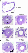
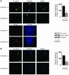
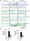
To determine whether endothelial Akt could affect vascular lesion formation, mutant mice with a constitutively active Akt transgene, which could be inducibly targeted to the vascular endothelium using the tet-off system (EC-Akt Tg mice), were generated. After withdrawal of doxycycline, EC-Akt Tg mice demonstrated increased endothelial-specific Akt activity and NO production. After blood flow cessation caused by carotid artery ligation, neointimal formation was attenuated in induced EC-Akt Tg mice compared with noninduced EC-Akt Tg mice and control littermates. To determine the role of eNOS in mediating these effects, mice were treated with N-nitro-L-arginine methyl ester (L-NAME). Neointimal formation was attenuated to a lesser extent in induced EC-Akt Tg mice treated with L-NAME, suggesting that some of the vascular protective effects were NO independent. Indeed, endothelial activation of Akt resulted in less EC apoptosis in ligated arteries. Immunostaining demonstrated decreased inflammatory and proliferative changes in induced EC-Akt Tg mice after vascular injury. These findings indicate that endothelial activation of Akt suppresses lesion formation via increased NO production, preservation of functional endothelial layer, and suppression of inflammatory and proliferative changes in the vascular wall. These results suggest that enhancing endothelial Akt activity alone could have therapeutic benefits after vascular injury.
Figures


Similar articles
-
Fas signaling induces Akt activation and upregulation of endothelial nitric oxide synthase expression.Hypertension. 2004 Apr;43(4):880-4. doi: 10.1161/01.HYP.0000120124.27641.03. Epub 2004 Feb 16. Hypertension. 2004. PMID: 14967838
-
Sphingosine-1-phosphate receptor 2 protects against anaphylactic shock through suppression of endothelial nitric oxide synthase in mice.J Allergy Clin Immunol. 2013 Nov;132(5):1205-1214.e9. doi: 10.1016/j.jaci.2013.07.026. Epub 2013 Sep 8. J Allergy Clin Immunol. 2013. PMID: 24021572
-
Overexpression of the β2AR gene improves function and re-endothelialization capacity of EPCs after arterial injury in nude mice.Stem Cell Res Ther. 2016 May 18;7(1):73. doi: 10.1186/s13287-016-0335-y. Stem Cell Res Ther. 2016. PMID: 27194135 Free PMC article.
-
Endothelial NO synthase overexpression inhibits lesion formation in mouse model of vascular remodeling.Arterioscler Thromb Vasc Biol. 2001 Feb;21(2):201-7. doi: 10.1161/01.atv.21.2.201. Arterioscler Thromb Vasc Biol. 2001. PMID: 11156853
-
Phosphodiesterase-5 inhibitor sildenafil preconditions adult cardiac myocytes against necrosis and apoptosis. Essential role of nitric oxide signaling.J Biol Chem. 2005 Apr 1;280(13):12944-55. doi: 10.1074/jbc.M404706200. Epub 2005 Jan 24. J Biol Chem. 2005. PMID: 15668244
Cited by
-
Multifaceted Mechanisms of Action of Metformin Which Have Been Unraveled One after Another in the Long History.Int J Mol Sci. 2021 Mar 5;22(5):2596. doi: 10.3390/ijms22052596. Int J Mol Sci. 2021. PMID: 33807522 Free PMC article. Review.
-
[Effect of glutaredoxin on oxidative stress of umbilical vein endothelial cell exposed to Porphyromonas gingivalis lipo- polysaccharide].Hua Xi Kou Qiang Yi Xue Za Zhi. 2015 Dec;33(6):613-6. doi: 10.7518/hxkq.2015.06.013. Hua Xi Kou Qiang Yi Xue Za Zhi. 2015. PMID: 27051955 Free PMC article. Chinese.
-
Gab2 (Grb2-Associated Binder2) Plays a Crucial Role in Inflammatory Signaling and Endothelial Dysfunction.Arterioscler Thromb Vasc Biol. 2021 Jun;41(6):1987-2005. doi: 10.1161/ATVBAHA.121.316153. Epub 2021 Apr 8. Arterioscler Thromb Vasc Biol. 2021. PMID: 33827252 Free PMC article.
-
Green tea polyphenol epigallocatechin gallate reduces endothelin-1 expression and secretion in vascular endothelial cells: roles for AMP-activated protein kinase, Akt, and FOXO1.Endocrinology. 2010 Jan;151(1):103-14. doi: 10.1210/en.2009-0997. Epub 2009 Nov 3. Endocrinology. 2010. PMID: 19887561 Free PMC article.
-
Loss of Akt activity increases circulating soluble endoglin release in preeclampsia: identification of inter-dependency between Akt-1 and heme oxygenase-1.Eur Heart J. 2012 May;33(9):1150-8. doi: 10.1093/eurheartj/ehr065. Epub 2011 Mar 16. Eur Heart J. 2012. PMID: 21411816 Free PMC article.
References
-
- Shimokawa H. Primary endothelial dysfunction: atherosclerosis. J. Mol. Cell. Cardiol. 1999;31:23–37. - PubMed
-
- Landmesser U, Hornig B, Drexler H. Endothelial function: a critical determinant in atherosclerosis? Circulation. 2004;109:II27–II33. - PubMed
-
- Bonetti PO, Lerman LO, Lerman A. Endothelial dysfunction: a marker of atherosclerotic risk. Arterioscler. Thromb. Vasc. Biol. 2003;23:168–175. - PubMed
-
- Melo LG, et al. Endothelium-targeted gene and cell-based therapies for cardiovascular disease. Arterioscler. Thromb. Vasc. Biol. 2004;24:1761–1774. - PubMed
Publication types
MeSH terms
Substances
Grants and funding
LinkOut - more resources
Full Text Sources
Other Literature Sources
Molecular Biology Databases
Miscellaneous

