. 2006 Feb;116(2):378-85.
doi: 10.1172/JCI25843.

Drusen deposits associated with aging and age-related macular degeneration contain nonfibrillar amyloid oligomers

Affiliations

Drusen deposits associated with aging and age-related macular degeneration contain nonfibrillar amyloid oligomers

Volker Luibl et al. J Clin Invest. 2006 Feb.

Abstract

Protein misfolding and aggregation are thought to underlie the pathogenesis of many amyloid diseases, such as Alzheimer and Parkinson diseases, whereby a stepwise protein misfolding process begins with the conversion of soluble protein monomers to prefibrillar oligomers and progresses to the formation of insoluble amyloid fibrils. Drusen are extracellular deposits found in aging eyes and in eyes afflicted with age-related macular degeneration (AMD). Recent characterizations of drusen have revealed protein components that are shared with amyloid deposits. However, characteristic amyloid fibrils have thus far not been identified in drusen. In this study, we tested the hypothesis that nonfibrillar oligomers may be a common link in amyloid diseases. Oligomers consisting of distinct amyloidogenic proteins and peptides can be detected by a recently developed antibody that is thought to recognize a common structure. Notably, oligomers exhibit cellular toxicity, which suggests that they play a role in the pathogenesis of neurodegenerative diseases. Through use of the anti-oligomer antibody, we came to observe the presence of nonfibrillar, toxic oligomers in drusen. Conversely, no reactivity was observed in age-matched control eyes without drusen. These results suggest that amyloid oligomers may be involved in drusen biogenesis and that similar protein misfolding processes may occur in AMD and amyloid diseases.


Figures
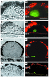
Figure 1
Figure 1
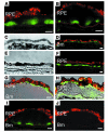
Immunolocalization of amyloidogenic oligomers in drusen by confocal laser microscopy. (A, C, E, and G) Differential interference contrast images. (B, D, F, and H) Confocal fluorescence images of amyloid oligomer cores (green, FITC channel). Drusen exhibit amyloid oligomer reactivity in the form of a core-like structure that accumulates centrally within drusen and in close proximity to the Bm. Autofluorescence of lipofuscin granules in the RPE cytoplasm is shown in red (Cy3 channel). (A and B) Anti-oligomer–specific antibody recognizes a spherical structure (∼15 μm) in a small druse (∼30 μm). (CF) Two larger drusen with centrally located core structure. (G and H) A very large macular soft druse from an 81-year-old female donor. Despite the difference in sizes and shapes of the drusen, the amyloid oligomer cores remain 10–15 μm in size. Scale bars: 10 μm.
Figure 2
Figure 2
Presence of amyloid oligomers in drusen and thickened Bm. Amyloid oligomer reactivity was visualized with fluorescein (green), and lipofuscin autofluorescence was visualized using the Cy3 channel (red). Multiple amyloid oligomer cores were sometimes observed in large drusen (A and B), as if a large druse may have formed from the fusion of several smaller drusen. The amyloid oligomer cores retained their size and relative positions within the druse and in proximity to the Bm. Within eyes that contained drusen, the oligomers occasionally accumulated above the Bm, in the form of basal linear (C and D) or basal laminar (EH) deposits, particularly in instances where the Bm appeared to be thickened. (H) Staining within RPE cells was also observed. C and D are differential interference contrast images of D and F, respectively. (I and J) Specificity of the antibody in cryosections is demonstrated in adjacent sections of a large druse. (I) Multiple amyloid oligomer cores were visualized through use of the anti-oligomer antibody. (J) Reactivity was eliminated when the primary antibody was preincubated with amyloid oligomers synthesized from the Aβ1–40 peptide. Scale bars: 10 μm.
Figure 3
Figure 3
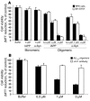
ELISA of retinal extracts using the A11 anti-oligomer antibody. (A) Increasing amounts of oligomers made from the Aβ1–40 peptide resulted in a dose-dependent response when incubated with the A11 anti-oligomer–specific antibody (filled circles). Little or no reactivity was observed when the Aβ1–40 oligomers were incubated without the primary antibody (open circles). (B) Dose-dependent reactivity was observed when the A11 antibody was incubated with increasing amounts of extract prepared from dissected drusen/RPE/Bm tissue from a 76-year-old male donor (filled circles). Little or no reactivity was observed when the primary antibody was omitted (open circles). Extracts prepared from the neural retina (filled triangles) of the same donor eye did not show a dose-dependent response when incubated with the A11 antibody. Dr, drusen.
Figure 4
Figure 4
Morphology of amyloid oligomer cores in drusen at higher magnification. (AC) Confocal micrographs of drusen. Amyloid oligomer cores are labeled with fluorescein (green), and lipofuscin autofluorescence in the RPE is visualized in red (Cy3 channel). (A) Amyloid oligomer cores seemed to consist of an aggregate of small vesicular structures (arrowheads) that increased in density toward the center. (B) Some of these vesicular structures appeared to extend toward the RPE with diminishing density (arrowheads). (C) Occasionally, the amyloid oligomer cores penetrated through the Bm and extended toward the choroid (arrowhead). (D) Ultrastructure of an amyloid oligomer core is depicted in an immunogold-labeled electron micrograph (inset), wherein gold particles decorate vesicular structures that are heterogeneous in size. The highest density of gold particles seen in D was from the region above the Bm (rectangle). Scale bars: 100 nm (D, inset). Magnification, ×3,000 (A), ×2,000 (BD).
Figure 5
Figure 5
Codistribution of amyloid oligomer cores and other known drusen components. (A, E, and I) Differential interference contrast images. (BD, FH, and JL) Confocal fluorescence images; amyloid oligomer cores were labeled with fluorescein (green). (A and B) Both antigens were present in a large druse, wherein the amyloid oligomer core was enveloped within the HLA-DR reactive region (labeled with Texas red). (C and D) At higher magnification, it is clear that the amyloid oligomer core and HLA-DR reactive subdomain did not colocalize in these drusen. (C) In one instance, the HLA-DR reactive region, perhaps reflecting a dendritic cell process, was observed as originating from the choroid (Ch), coming in close proximity to the Bm, and contacting the condensation of vesicular structures that represent the amyloid oligomer core. (D) In another instance, HLA-DR reactivity was observed as encompassing the choroid, the Bm, and the druse. Within the druse, HLA-DR reactivity appeared to surround the oligomer core, with no indication of colocalization. Similarly, no colocalization was observed with vitronectin (FH) or Aβ (JL), both labeled with Texas red (red). Lipofuscin autofluorescence within RPE is also visualized in the Cy3 channel (red). Scale bars: 10 μm. Magnification, ×250 (E, F, I, and J), ×1,500 (G), ×1,000 (H), and ×2,000 (K and L).
Figure 6
Figure 6
Toxicity of nonfibrillar amyloid oligomers to human primary RPE cells. (A) Amyloid oligomers were toxic to cultured SH-SY5Y human neuroblastoma cells (white bars) and human primary RPE cells (black bars). Oligomeric forms of IAPP and α-synuclein (α-Syn), but not soluble monomers, demonstrated toxicity. Cell viability was assessed by MTT reduction. (B) Increasing amounts of amyloid oligomers made from Aβ1–40 also showed a dose-dependent toxicity to cultured human primary RPE cells (black bars). This toxicity was largely blocked by adding equal molars of the A11 anti-oligomer antibody (white bars). Error bars represent SD; n = 3.

References

    1. Klein R, Peto T, Bird A, Vannewkirk MR. The epidemiology of age-related macular degeneration. Am. J. Ophthalmol. 2004;137:486–495. - PubMed
    1. Johnson PT, et al. Drusen-associated degeneration in the retina. Invest. Ophthalmol. Vis. Sci. 2003;44:4481–4488. - PubMed
    1. Abdelsalam A, Del Priore L, Zarbin MA. Drusen in age-related macular degeneration: pathogenesis, natural course, and laser photocoagulation-induced regression. Surv. Ophthalmol. 1999;44:1–29. - PubMed
    1. Crabb JW, et al. Drusen proteome analysis: an approach to the etiology of age-related macular degeneration. Proc. Natl. Acad. Sci. U. S. A. 2002;99:14682–14687. - PMC - PubMed
    1. Anderson DH, et al. Characterization of beta amyloid assemblies in drusen: the deposits associated with aging and age-related macular degeneration. Exp. Eye Res. 2004;78:243–256. - PubMed

Publication types