

Insulin's direct effects on the liver dominate the control of hepatic glucose production
- PMID: 16453026
- PMCID: PMC1359060
- DOI: 10.1172/JCI27073
Insulin's direct effects on the liver dominate the control of hepatic glucose production
Abstract
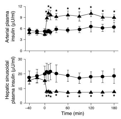
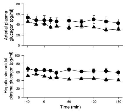
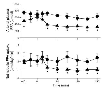
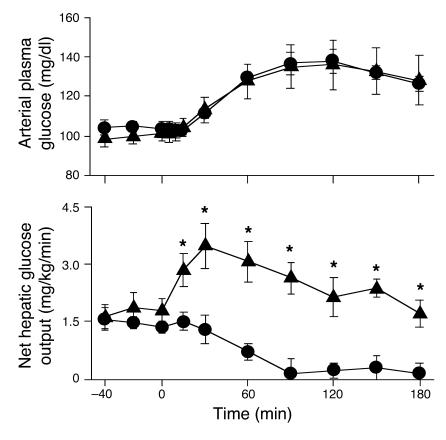
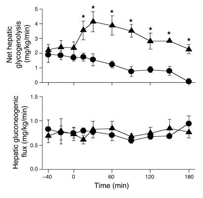
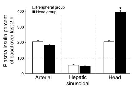
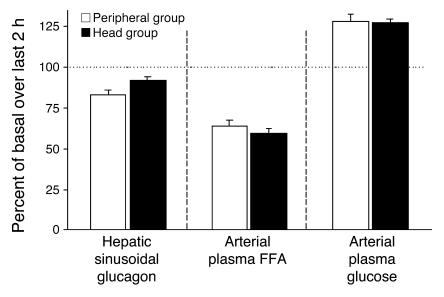
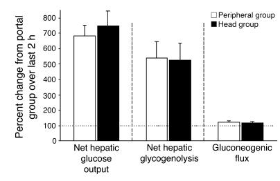
Insulin inhibits glucose production through both direct and indirect effects on the liver; however, considerable controversy exists regarding the relative importance of these effects. The first aim of this study was to determine which of these processes dominates the acute control of hepatic glucose production (HGP). Somatostatin and portal vein infusions of insulin and glucagon were used to clamp the pancreatic hormones at basal levels in the nondiabetic dog. After a basal sampling period, insulin infusion was switched from the portal vein to a peripheral vein. As a result, the arterial insulin level doubled and the hepatic sinusoidal insulin level was reduced by half. While the arterial plasma FFA level and net hepatic FFA uptake fell by 40-50%, net hepatic glucose output increased more than 2-fold and remained elevated compared with that in the control group. The second aim of this study was to determine the effect of a 4-fold rise in head insulin on HGP during peripheral hyperinsulinemia and hepatic insulin deficiency. Sensitivity of the liver was not enhanced by increased insulin delivery to the head. Thus, this study demonstrates that the direct effects of insulin dominate the acute regulation of HGP in the normal dog.
Figures
Comment in
-
Insulin's effect on the liver: "direct or indirect?" continues to be the question.J Clin Invest. 2006 Feb;116(2):302-4. doi: 10.1172/JCI27743. J Clin Invest. 2006. PMID: 16453016 Free PMC article.
References
-
- DeFronzo RA, Bonadonna RC, Ferrannini E. Pathogenesis of NIDDM. A balanced overview. Diabetes Care. 1992;15:318–368. - PubMed
-
- Cherrington AD, Edgerton D, Sindelar DK. The direct and indirect effects of insulin on hepatic glucose production in vivo. Diabetologia. 1998;41:987–996. - PubMed
-
- Claus TH, Pilkis SJ. Regulation by insulin of gluconeogenesis in isolated rat hepatocytes. Biochim. Biophys. Acta. 1976;421:246–262. - PubMed
-
- Marks JS, Botelho LH. Synergistic inhibition of glucagon-induced effects on hepatic glucose metabolism in the presence of insulin and a cAMP antagonist. J. Biol. Chem. 1986;261:15895–15899. - PubMed
-
- Sindelar DK, Balcom JH, Chu CA, Neal DW, Cherrington AD. A comparison of the effects of selective increases in peripheral or portal insulin on hepatic glucose production in the conscious dog. Diabetes. 1996;45:1594–1604. - PubMed
Publication types
MeSH terms
Substances
Grants and funding
LinkOut - more resources
Full Text Sources
Medical
Research Materials
