. 2006 Mar 1;193(5):625-33.
doi: 10.1086/500148. Epub 2006 Feb 2.

Efficient neutralization of anthrax toxin by chimpanzee monoclonal antibodies against protective antigen

Affiliations

Efficient neutralization of anthrax toxin by chimpanzee monoclonal antibodies against protective antigen

Zhaochun Chen et al. J Infect Dis. .

Abstract

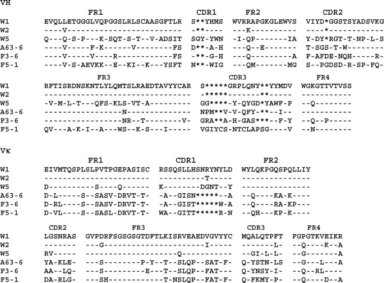
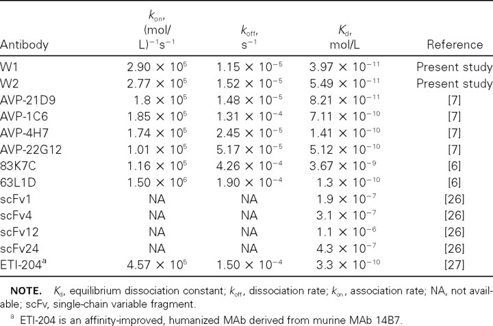
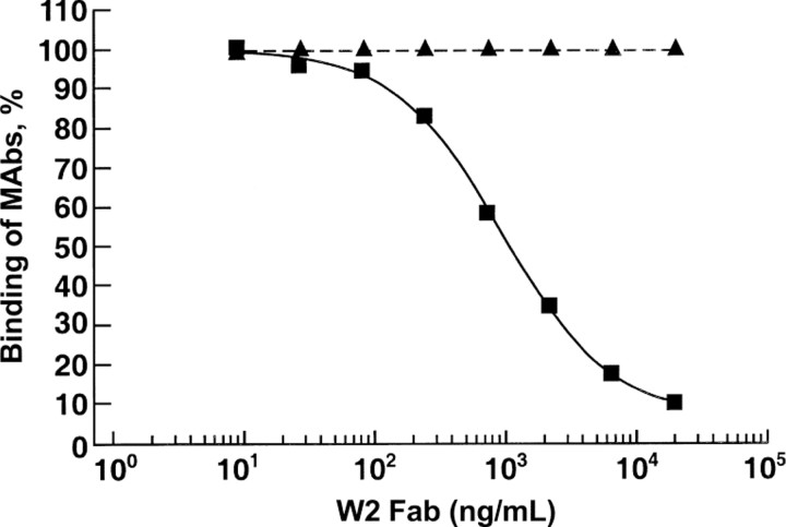
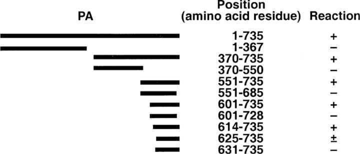
Four single-chain variable fragments (scFvs) against protective antigen (PA) and 2 scFvs against lethal factor (LF) of anthrax were isolated from a phage display library generated from immunized chimpanzees. Only 2 scFvs recognizing PA (W1 and W2) neutralized the cytotoxicity of lethal toxin in a macrophage lysis assay. Full-length immunoglobulin G (IgG) of W1 and W2 efficiently protected rats from anthrax toxin challenge. The epitope recognized by W1 and W2 was conformational and was formed by C-terminal amino acids 614-735 of PA. W1 and W2 each bound to PA with an equilibrium dissociation constant of 4x10-11 mol/L to 5x10(-11) mol/L, which is an affinity that is 20-100-fold higher than that for the interaction of the receptor and PA. W1 and W2 inhibited the binding of PA to the receptor, suggesting that this was the mechanism of protection. These data suggest that W1 and W2 chimpanzee monoclonal antibodies may serve as PA entry inhibitors for use in the emergency prophylaxis against and treatment of anthrax.


Figures

Figure 1
Figure 1
Alignment of the deduced amino acid sequences of the variable domains of the heavy chain (VH) and κ chain (Vκ). Substitutions relative to W1 are expressed as single letters denoting amino acids. Dashes denote identical residues, and asterisks denote the absence of corresponding residues relative to the longest sequence. Complementarity-determining regions (CDR1–3) and framework regions (FR1–4) are shown above the sequence alignments
Table 1
Table 1
Human immunoglobulin germline genes most closely related to the chimpanzee heavy chain (VH) and κ light chain (Vκ) of various anti-anthrax protective antigen (PA) and lethal factor (LF) antibodies
Figure 2
Figure 2
ELISA titration of anti–protective antigen (PA) (A) and anti–lethal factor (LF) (B) single-chain variable fragments (scFvs). Recombinant PA or LF or unrelated proteins, bovine serum albumin (BSA), thyroglobulin, lysozyme, and phosphorylase b were used to coat the wells of an ELISA plate. Wells then were incubated with various dilutions of scFvs. Bound scFv was detected by the addition of peroxidase-conjugated anti-His antibody, followed by tetramethylbenzidine substrate. The anti-PA and anti-LF scFvs did not bind to the unrelated proteins; only BSA is shown as an example. OD, optical density measured at 450 nm
Figure 3
Figure 3
In vitro neutralization assay. Anti–protective antigen (PA) IgG was mixed with anthrax toxin and was incubated for 1 h at 37°C. The mixture was added to RAW264.7 cells in a 96-well plate and was incubated for 4 h at 37°C. After washing, the cells were stained with MTT dye, and then lysis was performed in a solution containing 0.5% SDS in 90% isopropanol and 0.05 mol/L HCl. The plate was read at an optical density measured at 570 nm, and the cell survival was calculated relative to untreated cells. Results were plotted and analyzed using Prism software (version 4; Graphpad Software). EC50, effective concentration for 50% neutralization. ▪, W1; ▾, W2; □, 14B7
Table 2
Table 2
Affinity of anti-anthrax protective antigen (PA) W1 and W2 antibodies, compared with other human anti-PA monoclonal antibodies (MAbs)
Figure 4
Figure 4
Competitive ELISA. Recombinant protective antigen (PA) was coated onto the wells of an ELISA plate. Wells then were incubated with anti-PA W2 Fab at the concentrations indicated. After incubation for 1 h at room temperature, anti-PA W2 Fab was removed from the wells, and mouse anti-PA monoclonal antibodies (MAbs) 14B7 and 2D3 [16, 23] were added to the wells. Bound MAbs were detected by the addition of peroxidase-conjugated anti–mouse antibody, followed by tetramethylbenzidine substrate. The binding to PA was calculated by dividing the optical density value in the absence of W2 by the optical density value in the presence of W2. ▪, 14B7; ▴, 2D3
Figure 5
Figure 5
Summary of epitope mapping of anti–protective antigen (PA) W2 antibody by radioimmunoprecipitation assay. 35S-labeled PA peptides, prepared in vitro, were incubated with anti-PA W2. The immune complexes were captured by protein G–coupled agarose beads and were separated by SDS-PAGE. The PA peptide was detected by exposing the dried gel to an x-ray film. Numbers denote the starting and ending amino acids. The peptides that reacted with antibody and, hence, were detected on an x-ray film were considered to be positive (+). Faint intensity of the band on an x-ray film denoted a partial reaction (±)
Figure 6
Figure 6
Inhibition of the binding of protective antigen (PA) to RAW264.7 cells by preincubation of toxin with monoclonal antibodies. PA at a concentration of 6 nmol/L (500 ng/mL) was incubated with anti-PA 14B7 or W2 antibodies at a molar ratio of 1:1 or 1:10 for 5 min. The mixture was added to RAW264.7 cells and was incubated for 20 min at 37°C. After the cells were washed and lysed, separation by SDS-PAGE was performed. The proteins were transferred to a membrane and were probed with anti-PA polyclonal antibody
Table 3
Table 3
Neutralization of protective antigen (PA) of anthrax toxin by W2 in rats
Table 4
Table 4
Duration of neutralization of protective antigen (PA) of anthrax toxin by W1 and W2 in rats

Similar articles

Cited by

References

    1. Jernigan JA, Stephens DS, Ashford DA, et al. Bioterrorism‐related inhalational anthrax: the first 10 cases reported in the United States. Emerg Infect Dis. 2001;7:933–44. - PMC - PubMed
    1. Collier RJ, Young JA. Anthrax toxin. Annu Rev Cell Dev Biol. 2003;19:45–70. - PubMed
    1. Little SF, Ivins BE, Fellows PF, Friedlander AM. Passive protection by polyclonal antibodies against Bacillus anthracis infection in guinea pigs. Infect Immun. 1997;65:5171–5. - PMC - PubMed
    1. Kobiler D, Gozes Y, Rosenberg H, Marcus D, Reuveny S, Altboum Z. Efficiency of protection of guinea pigs against infection with Bacillus anthracis spores by passive immunization. Infect Immun. 2002;70:544–60. - PMC - PubMed
    1. Maynard JA, Maassen CB, Leppla SH, et al. Protection against anthrax toxin by recombinant antibody fragments correlates with antigen affinity. Nat Biotechnol. 2002;20:597–601. - PubMed

Publication types

MeSH terms