

Review
doi: 10.1136/bmj.332.7536.285.
Wound assessment
Affiliations
- PMID: 16455730
- PMCID: PMC1360405
- DOI: 10.1136/bmj.332.7536.285
Item in Clipboard
Review
Wound assessment
BMJ.
.
No abstract available
Figures
Wounds are not just skin deep, and accurate assessment is an essential part of treatment
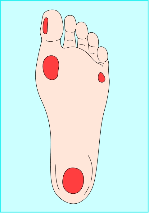
Areas of abnormal pressure distribution in the diabetic foot. Plantar ulcers are most commonly seen under the hallux, on the first and fifth metatarsal heads, and under the heel
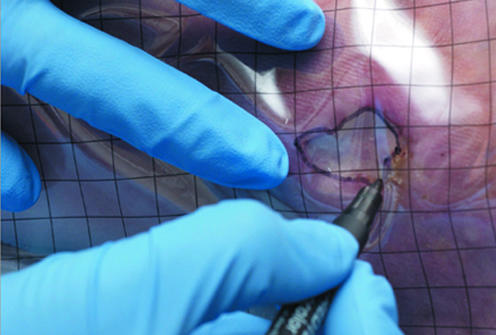
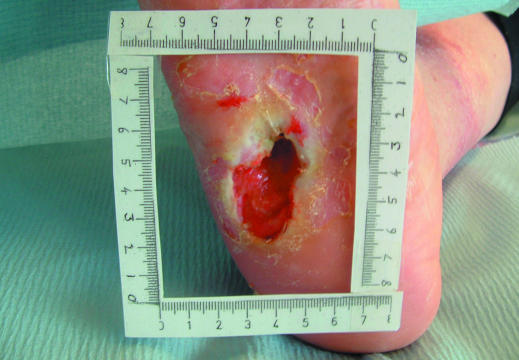
Tracing a wound for measurement and measuring a wound
Tracing a wound for measurement and measuring a wound
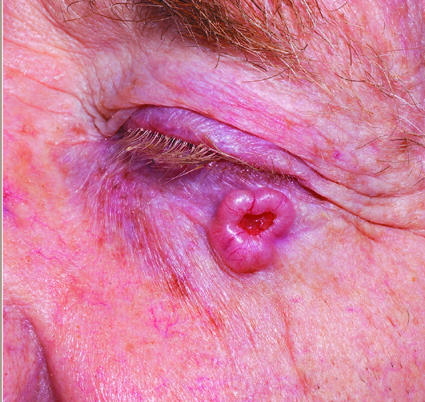
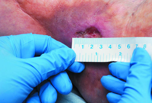
Left: Basal cell carcinoma with rolled edges. Right: Lymphoma presenting as groin ulceration
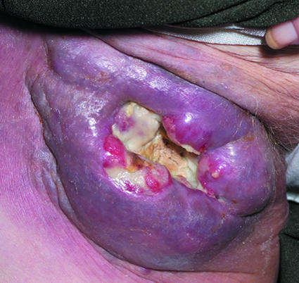
Left: Basal cell carcinoma with rolled edges. Right: Lymphoma presenting as groin ulceration
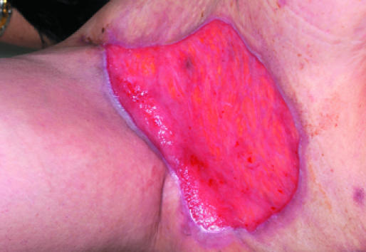
Left: Healthy granulation tissue in a hidradenitis suppurativa excision wound. Right: Unhealthy granulation tissue in a venous leg ulcer
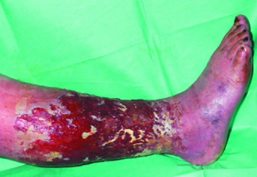
Left: Healthy granulation tissue in a hidradenitis suppurativa excision wound. Right: Unhealthy granulation tissue in a venous leg ulcer
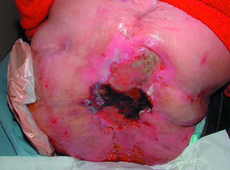
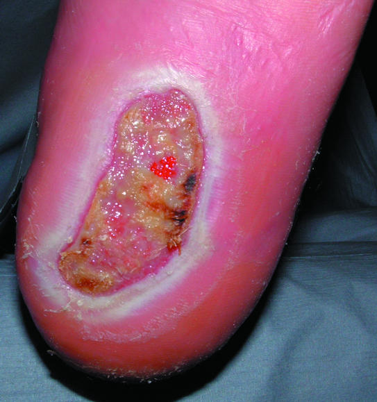
Top: Necrotic tissue (black areas) in a pressure ulcer. Bottom: Slough at the base of a pressure ulcer. Right: Eschar covering a heel pressure ulcer
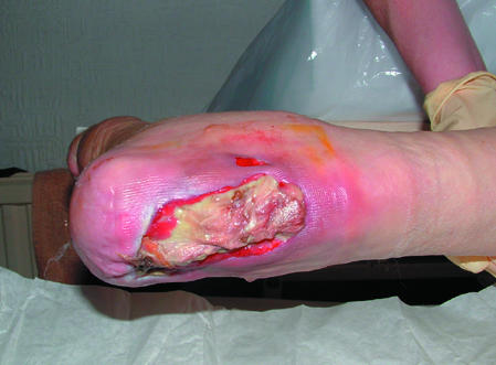
Top: Necrotic tissue (black areas) in a pressure ulcer. Bottom: Slough at the base of a pressure ulcer. Right: Eschar covering a heel pressure ulcer
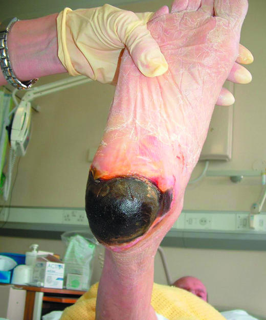
Top: Necrotic tissue (black areas) in a pressure ulcer. Bottom: Slough at the base of a pressure ulcer. Right: Eschar covering a heel pressure ulcer
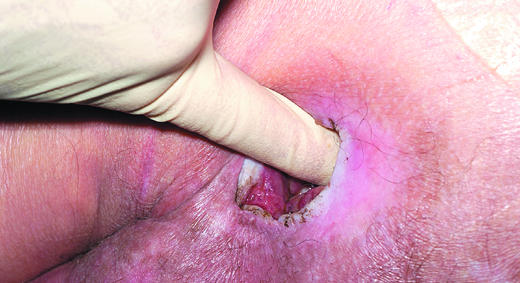
Left: Digital examination of a wound. Right: Examining a wound with a probe

Left: Digital examination of a wound. Right: Examining a wound with a probe
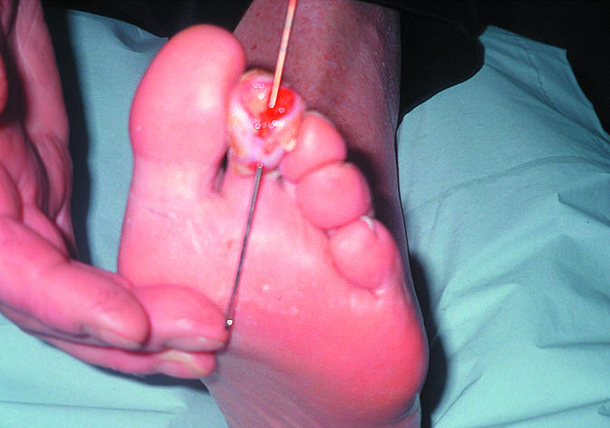
Fistula in a diabetic foot ulcer
Maceration of the skin surrounding a diabetic foot ulcer
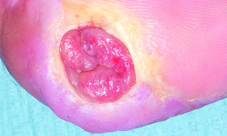
Overgranulation may be a sign of infection or non-healing
References
-
- Lazarus GS, Cooper DM, Knighton DR, Margolis DJ, Pecoraro RE, Rodeheaver G, et al. Definitions and guidelines for assessment of wounds and evaluation of healing. Arch Dermatol 1994;130: 489-93. - PubMed
-
- Izadi K, Ganchi P. Chronic wounds. Clin Plast Surg 2005;32: 209-22. - PubMed
-
- Falanga V, Phillips TJ, Harding KG, Moy RL, Peerson LJ, eds. Text atlas of wound management. London: Martin Dunitz, 2000.
Publication types
MeSH terms
LinkOut - more resources
Full Text Sources
Other Literature Sources
Medical