. 2006 Feb 28;103(9):3244-9.
doi: 10.1073/pnas.0510748103. Epub 2006 Feb 2.

Anthrax lethal factor and edema factor act on conserved targets in Drosophila

Affiliations

Anthrax lethal factor and edema factor act on conserved targets in Drosophila

Annabel Guichard et al. Proc Natl Acad Sci U S A. .

Abstract

Many bacterial toxins act on conserved components of essential host-signaling pathways. One consequence of this conservation is that genetic model organisms such as Drosophila melanogaster can be used for analyzing the mechanism of toxin action. In this study, we characterize the activities of two anthrax virulence factors, lethal factor (LF) and edema factor, in transgenic Drosophila. LF is a zinc metalloprotease that cleaves and inactivates most human mitogen-activated protein kinase (MAPK) kinases (MAPKKs). We found that LF similarly cleaves the Drosophila MAPK kinases Hemipterous (Hep) and Licorne in vitro. Consistent with these observations, expression of LF in Drosophila inhibited the Hep/c-Jun N-terminal kinase pathway during embryonic dorsal closure and the related process of adult thoracic closure. Epistasis experiments confirmed that LF acts at the level of Hep. We also found that LF inhibits Ras/MAPK signaling during wing development and that LF acts upstream of MAPK and downstream of Raf, consistent with LF acting at the level of Dsor. In addition, we found that edema factor, a potent adenylate cyclase, inhibits the hh pathway during wing development, consistent with the known role of cAMP-dependent PKA in suppressing the Hedgehog response. These results demonstrate that anthrax toxins function in Drosophila as they do in mammalian cells and open the way to using Drosophila as a multicellular host system for studying the in vivo function of diverse toxins and virulence factors.


Conflict of interest statement

Conflict of interest statement: E.B. holds shares in NovaScape Sciences.

Figures
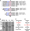
Fig. 1.
Fig. 1.
In vitro cleavage of Drosophila MAPKKs by anthrax LF. (A) A multiple protein sequence alignment of known LF cleavage sites in human MAPKKs and cognate sequences in Drosophila MAPKKs. Conserved residues defining the cleavage motif are denoted in color (blue, basic residues; red, hydrophobic residues). LF cleavage sites are indicated by arrows. Amino acid residues flanking known cleavage sites are in parentheses. (B) 35S-labeled MAPKK proteins were synthesized in vitro and incubated with LF (250 ng) for 1 h. MAPKK cleavage was analyzed by SDS/PAGE and autoradiography. (C) Schematic diagram of JNK pathway components in Drosophila and mammals (parentheses) examined in this study. (D) Schematic diagram of RTK/RAS pathway components. Mammalian homologs appear in parentheses.
Fig. 2.
Fig. 2.
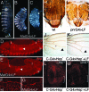
LF inhibits Hep/JNK signaling in Drosophila. (AC) Cuticle preparations of Drosophila wild-type (wt) (A) or MatGAL4>LF (B and C) embryos. LF-expressing embryos are similar to Hep/JNK pathway mutant embryos (21, 30). GAL4 drivers indicated in labels in this and subsequent figures are abbreviated as G4. Among embryos expressing high levels of LF that have dorsal closure defects the various phenotypic categories occur at the following respective frequencies (n = 54): U-shaped (46%), U-shaped with head cuticle defects and/or dorsal holes (42%), and severe cuticle phenotype (12%). In C the arrow indicates an anterior dorsal hole in the cuticle. (D and E) in situ hybridization of Drosophila embryos using a dpp antisense probe. Brackets indicate leading-edge cells, which express dpp in wild-type embryos (D), but fail to do so in MatGAL4>LF embryos (E). Arrows point to dpp-expressing midgut cells, and the asterisk indicates a lateral stripe of dpp expression, independent of JNK regulation. (F and G) High-magnification views of F-actin in embryos stained with Alexa Fluor 555-coupled phalloidin. Elongated leading-edge cells have a sharp and regular F-actin front in wild-type (wt) embryos (F), whereas in MatGAL4>LF embryos (G) the F-actin front is irregular and discontinuous. (H and I) Thoraxes dissected from wild-type (wt) (H) and pnrGAL4>LF (I) adults. LF-expressing individuals display a dorsal cleft phenotype typical of Hep viable mutants (33). (J and K) Proximal portions of adult wings. (J) A C-GAL4>Hep-CA (activated Hep) wing. (K) A C-GAL4>Hep-CA+LF wing, in which LF suppresses the phenotype induced by Hep-CA. (L and M) Third-instar imaginal discs stained with an anti-activated JNK antibody. (L) A C-GAL4>Hep-CA disc. Bracket indicates the posterior domain of GAL4 expression. (M) A C-GAL4>Hep-CA+LF disc showing reduced JNK activation caused by LF.
Fig. 3.
Fig. 3.
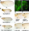
LF inhibits Dsor/MAPK signaling. (AD) Adult wings. Stg-wingGAL4>LF refers to males with three copies of the UAS-LF construct driven by the MS-1096 ubiquitous wing driver at 29°C. The small wing vein phenotype (B) has nearly 100% penetrance, and ≈50% of these individuals also have vein truncations (not present in the wing shown in B). Mod-wingGAL4 refers to the MS1096GAL4 driving expression of three copies of the UAS-LF construct in females at 25°C (A). (D) The Dsor1 allele used is Dsor1rl. Arrow points at a truncated L5 vein. (E and F) Third-instar imaginal discs stained with an anti-di-phosphoMAPK antibody. (E) A wild-type (wt) disc, showing localized MAPK activation in the margin and vein primordial. (F) An MS1096>LF disc, in which LF suppresses MAPK activation. (G) An MS1096>UAS-Sem wing, in which the activated allele Sem of the rolled MAPK causes ectopic veins (arrows). This phenotype is not suppressed by coexpression of LF (H). (I and J) Medial portions of adult wings, showing ectopic veins (arrows) in C-GAL4>Rafgof wings (I) and suppression of this phenotype by LF (J).
Fig. 4.
Fig. 4.
EF inhibits PKA-dependent Hh signaling. (AE) Adult wings. Brackets indicate the distance between longitudinal veins L3 and L4, which is reduced in dpp-discGAL4>EF wings (B). This phenotype is clearly enhanced in hh heterozygous mutants (C). (D) A dppGAL4>PKAr wing, in which expression of the regulatory subunit of PKA causes sublethality and severe patterning defects. This phenotype is almost completely suppressed by coexpression with EF (E). (FI) Anterior legs of adult males. Expression of the regulatory subunit of PKA causes malformation of legs (G). This phenotype is also suppressed by coexpression with EF (I). EF expression with other GAL4 drivers causes lethality (apGAL4, ptcGAL4, and dppGAL4 at 25°C), strong composite wing phenotypes (71BGAL4, 1348GAL4, scaGAL4, MS1096GAL4, and vgGAL4), and small or rough eyes (eyeGAL4 and GMRGAL4, respectively). wt, wild type.

Comment in

  • Fighting anthrax with flies.
    Giagtzoglou N, Bellen HJ. Giagtzoglou N, et al. Proc Natl Acad Sci U S A. 2006 Feb 28;103(9):3013-4. doi: 10.1073/pnas.0600477103. Epub 2006 Feb 21. Proc Natl Acad Sci U S A. 2006. PMID: 16492749 Free PMC article. No abstract available.

Similar articles

Cited by

References

    1. Reiter L. T., Potocki L., Chien S., Gribskov M., Bier E. Genome Res. 2001;11:1114–1125. - PMC - PubMed
    1. Bier E. Nat. Rev. Genet. 2005;6:9–23. - PubMed
    1. Bier E., McGinnis W. In: Molecular Basis of Inborn Errors of Development. Epstein C. J., Erikson R. P., Wynshaw-Boris A., editors. New York: Oxford Univ. Press; 2004. pp. 25–45.
    1. Beutler B. Mol. Immunol. 2004;40:845–859. - PubMed
    1. Collier R. J., Young J. A. Annu. Rev. Cell Dev. Biol. 2003;19:45–70. - PubMed

Publication types

MeSH terms

LinkOut - more resources