

doi: 10.1128/JCM.44.2.583-585.2006.
Use of different stains for microscopic evaluation of corneal scrapings for diagnosis of microsporidial keratitis
Affiliations
- PMID: 16455916
- PMCID: PMC1392708
- DOI: 10.1128/JCM.44.2.583-585.2006
Item in Clipboard
Use of different stains for microscopic evaluation of corneal scrapings for diagnosis of microsporidial keratitis
J Clin Microbiol.
2006 Feb.
Abstract
Retrospective evaluation of potassium hydroxide plus calcofluor white (KOH+CFW), Gram, Giemsa, and modified Ziehl-Neelsen (1% H(2)SO(4), cold) stains for the detection of microsporidia in corneal scrapings from 30 patients showed KOH+CFW and acid-fast stains to be most efficient (29/30 [96.7%] and 28/30 [93.3%], respectively) in the diagnosis of microsporidial keratitis.
Figures
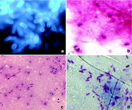
Microsporidal spores as observed under various stains on corneal scrapings. (a) KOH+CFW stain (magnification, ×1,000). Organisms were seen as bright turquoise to white oval bodies, often clustered in groups, against a relatively dark background. The spores displayed variable fluorescence intensities. Depending on the orientation of the microsporidia, the anterior end appeared concave. (b) Gram stain (magnification, ×1,000). Spores appeared ovoid and refractile and bright purple, resembling gram-positive organisms. The spores were scattered or highly clustered within the cytoplasm of occasional epithelial cells. Microsporidial spores show a dark staining belt girding them either diagonally or equatorially. (c) Giemsa stain (magnification, ×1,000). This stain is not taken up by the cell wall, and only the cytoplasm gets stained. The spores appear smaller than those in the other stains. There was also poor differentiation from other bacteria and debris. The darkly stained belt could be identified in 18/30 cases, aiding preliminary diagnosis. (d) Modified Ziehl-Neelsen stain (magnification, ×1,000). Except for two, all cases of microsporidial spores were acid fast (1% H2SO4). The acid-fast spores appeared bright red on a blue background, and a posterior vacuole and central diagonal strip within the spores were often visible. Bacteria and other cell debris appeared blue, owing to methylene blue counterstain.
Similar articles
-
Use of potassium hydroxide, Giemsa and calcofluor white staining techniques in the microscopic evaluation of corneal scrapings for diagnosis of fungal keratitis.J Int Med Res. 2010;38(6):1961-7. doi: 10.1177/147323001003800609. J Int Med Res. 2010. PMID: 21226999
-
Diagnosis of microsporidial keratitis by confocal microscopy and the chromatrope stain.Am J Ophthalmol. 1996 Jan;121(1):89-91. doi: 10.1016/s0002-9394(14)70538-0. Am J Ophthalmol. 1996. PMID: 8554085
-
Masked comparison of trypan blue stain and potassium hydroxide with calcofluor white stain in the microscopic examination of corneal scrapings for the diagnosis of microbial keratitis.Indian J Ophthalmol. 2021 Sep;69(9):2457-2460. doi: 10.4103/ijo.IJO_3685_20. Indian J Ophthalmol. 2021. PMID: 34427244 Free PMC article.
-
Parasitic corneal infections.Int Ophthalmol Clin. 1998 Fall;38(4):179-82. doi: 10.1097/00004397-199803840-00016. Int Ophthalmol Clin. 1998. PMID: 10081733 Review. No abstract available.
-
Microsporidial keratitis: need for increased awareness.Surv Ophthalmol. 2011 Jan-Feb;56(1):1-22. doi: 10.1016/j.survophthal.2010.03.006. Epub 2010 Nov 11. Surv Ophthalmol. 2011. PMID: 21071051 Review.
Cited by
-
Evaluating the accuracy and diagnostic value of CFW and a new fluorescent reagents, fluorescent brightener 85, for the diagnosis of vulvovaginal candidiasis.J Clin Lab Anal. 2021 Aug;35(8):e23891. doi: 10.1002/jcla.23891. Epub 2021 Jul 12. J Clin Lab Anal. 2021. PMID: 34251053 Free PMC article.
-
Comparison of fungal fluorescent staining and ITS rDNA PCR-based sequencing with conventional methods for the diagnosis of onychomycosis.J Eur Acad Dermatol Venereol. 2018 Jun;32(6):1017-1021. doi: 10.1111/jdv.14843. Epub 2018 Feb 27. J Eur Acad Dermatol Venereol. 2018. PMID: 29405481 Free PMC article.
-
Microsporidial Stromal Keratitis in Post-Keratoplasty Eyes.J Clin Med. 2023 May 27;12(11):3706. doi: 10.3390/jcm12113706. J Clin Med. 2023. PMID: 37297901 Free PMC article.
-
Pseudoloma neurophilia infections in zebrafish Danio rerio: effects of stress on survival, growth, and reproduction.Dis Aquat Organ. 2009 Dec 22;88(1):69-84. doi: 10.3354/dao02145. Dis Aquat Organ. 2009. PMID: 20183967 Free PMC article.
-
A study on the accuracy of a new fluorescent detection method for vaginal fungi.BMC Womens Health. 2022 Dec 30;22(1):559. doi: 10.1186/s12905-022-02151-9. BMC Womens Health. 2022. PMID: 36585693 Free PMC article.
References
-
- Awadalla, H. N., I. F. el Naga, M. M. el-Temsahi, and A. Y. Negm. 1998. Detection of microsporidia by different staining techniques. J. Egypt. Soc. Parasitol. 28:729-738. - PubMed
-
- Canning, E. U., and W. S. Hollister. 1987. Microsporidia of mammals—widespread pathogens or opportunistic curiosities? Parasitol. Today 9:267-273. - PubMed
-
- Chan, C. M., J. T. Theng, L. Li, and D. T. Tan. 2003. Microsporidial keratoconjunctivitis in healthy individuals: a case series. Ophthalmology 110:1420-1425. - PubMed
-
- Davis, R., R. Font, M. Keisler, and J. Shadduck. 1990. Corneal microsporidiosis: a case report including ultrastructural observations. Ophthalmology 97:953-957. - PubMed
-
- Joste, N. E., P. E. Sax, and W. S. Pieciak. 1999. Cytologic detection of microsporidia spores in bile. A comparison of stains. Acta Cytol. 43:98-103. - PubMed
Publication types
MeSH terms
Substances
LinkOut - more resources
Full Text Sources
