. 2006 May 15;107(10):3902-6.
doi: 10.1182/blood-2005-09-3687. Epub 2006 Feb 2.

Glycoprotein VI-dependent and -independent pathways of thrombus formation in vivo

Affiliations

Glycoprotein VI-dependent and -independent pathways of thrombus formation in vivo

Christophe Dubois et al. Blood. .

Abstract

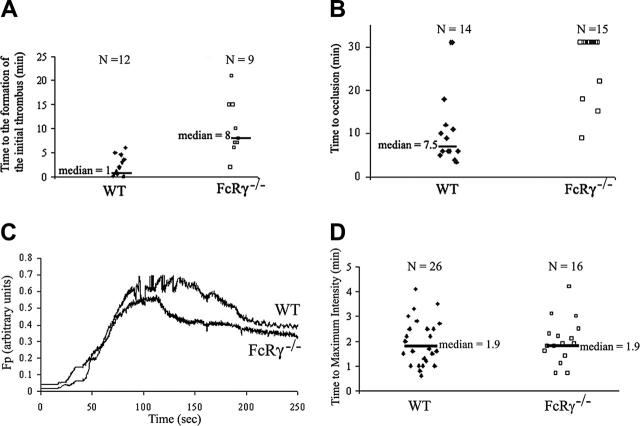
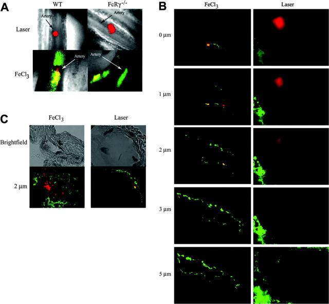
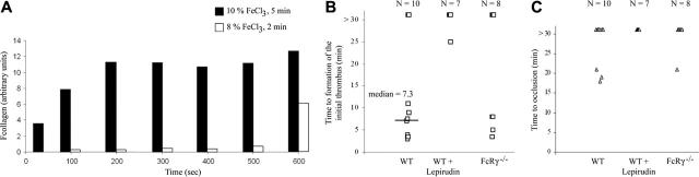
The role of the collagen receptor glycoprotein VI (GPVI) in arteriolar thrombus formation was studied in FcRgamma-null mice (FcRgamma(-/-)) lacking platelet surface GPVI. Thrombi were induced with severe or mild FeCl(3) injury. Collagen exposure was significantly delayed and diminished in mild compared with severe FeCl(3) injury. Times to initial thrombus formation and vessel occlusion were delayed in FcRgamma(-/-) compared with wild-type mice after severe injury. Platelet accumulation in wild-type mice was decreased after mild compared with severe injury. However, there was little difference between platelet accumulation after severe or mild injury in FcRgamma(-/-). These data indicate a significant role for GPVI in FeCl(3)-induced thrombus formation. Pretreatment of wild-type mice with lepirudin further impaired mild FeCl(3)-induced thrombus formation, demonstrating a role for thrombin. Laser-induced thrombus formation in wild-type and FcRgamma(-/-) was comparable. Collagen exposure to circulating blood was undetectable after laser injury. Normalized for thrombus size, thrombus-associated tissue factor was 5-fold higher in laser-induced thrombi than in severe FeCl(3)-induced thrombi. Thus, platelet activation by thrombin appears to be more important after laser injury than platelet activation by GPVI-collagen. It may thus be important when considering targets for antithrombotic therapy to use multiple animal models with diverse pathways to thrombus formation.


Figures

Figure 1.
Figure 1.
Thrombus formation in FcRγ-null mice and wild-type mice in the FeCl3 and laser-induced thrombosis models. Platelets were labeled by infusion of Alexa 660–conjugated rat anti–mouse CD41 antibody (250 ng/g body weight). (A-B) Injuries were induced with 10% FeCl3 for 5 minutes in wild-type and FcRγ-null mice. (A) The time to the formation of an initial thrombus (WT, n = 12; FcRγ–/–, n = 9). (B) The time to vessel occlusion (WT mice, n = 14; FcRγ–/–, n = 15) are reported and the median times calculated. (C-D) Laser-induced injuries in cremaster arterioles of wild-type and FcRγ-null mice. (C) Median integrated platelet fluorescence intensity for multiple thrombi of each genotype: WT (26 thrombi in 3 mice); FcRγ–/– (16 thrombi in 3 mice). Fluorescence of platelets in arbitrary units is presented as a function of time. (D) For each thrombus formed, the time to the maximum platelet accumulation into the growing thrombus is reported and the calculated median time indicated.
Figure 2.
Figure 2.
Detection of collagen type I and tissue factor in thrombi after FeCl3 or laser-induced injury. (A) Injuries in wild-type or FcRγ-null mice were induced by application of 10% FeCl3 for 5 minutes or by laser. Platelets were labeled by injection of Alexa 660–conjugated rat anti-CD41 antibody into the circulation prior to injury (red pseudocolor). Collagen was labeled by injection of a rabbit anti–mouse collagen type I antibody (1 μg/g body weight) plus an Alexa 488–conjugated goat anti–rabbit antibody (1 μg/g body weight) prior to injury (green pseudocolor). Merge of platelets and collagen, yellow pseudocolor. (B-C) Cremaster muscles injured by FeCl3 or by laser were isolated and sectioned. Image sections of a thrombus were acquired at 0.5-μm intervals across the vessel diameter perpendicular to the long axis of the vessel by incrementally altering the working distance between the microscope objective and the speciment with a piezo-electric driver. (B) Tissue sections were stained with Alexa 660 anti–mouse CD41 antibody, rabbit anti–mouse collagen type I antibody, and an Alexa 488–conjugated goat antirabbit antibody. Platelets are indicated by red pseudocolor; collagen, green pseudocolor; and the merge, yellow pseudocolor. Pictures shown are representative of 4 independent experiments. (C) Tissue sections were stained with Alexa 660–conjugated anti–mouse CD41 antibody, rabbit anti–mouse tissue factor (TF) antibody, and Alexa 488–conjugated goat antirabbit antibody. Platelets are indicated by red pseudocolor; tissue factor, green pseudocolor; and the merge, yellow pseudocolor. Pictures shown are representative of 4 independent experiments.
Figure 3.
Figure 3.
Collagen exposure after injury at varying FeCl3 concentrations and effect of lepirudin on thrombus formation in wild-type mice. (A) Injuries in wild-type mice (WT) were induced by application of 10% or 8% FeCl3 for 5 or 2 minutes, respectively. Collagen exposed to the blood circulation was labeled by injection of a rabbit anti–mouse collagen type I antibody (1 μg/g body weight) plus Alexa 488–conjugated goat antirabbit antibody (1μg/g body weight). Integrated fluorescence intensity of collagen at several time points after injury is indicated for individual thrombi under the 2 conditions (n = 5 for each condition). (B) Local administration of an 8% solution of FeCl3 for 2 minutes to the adventitial surface of the mesentery was used to induce vascular injury in wild-type mice. Mice were treated with 5 μg/g body weight of lepirudin prior to vessel injury where indicated. For each thrombus the time to the formation of an initial thrombus is reported and the median time indicated (n = 10). (C) For each thrombus in panel B the time to vessel occlusion is reported. Most vessels did not occlude during the 30 minutes of the experiment.

Similar articles

Cited by

References

    1. Baumgartner HR, Tschopp TB, Weiss HJ. Platelet interaction with collagen fibrils in flowing blood, II: impaired adhesion-aggregation in bleeding disorders: a comparison with subendothelium. Thromb Haemost. 1977;37: 17-28. - PubMed
    1. Meyer D, Baumgartner HR. Role of von Willebrand factor in platelet adhesion to the subendothelium. Br J Haematol. 1983;54: 1-9. - PubMed
    1. Santoro SA. Identification of a 160,000 dalton platelet membrane protein that mediates the initial divalent cation-dependent adhesion of platelets to collagen. Cell. 1986;46: 913-920. - PubMed
    1. Moroi M, Jung SM, Okuma M, Shinmyozu K. A patient with platelets deficient in glycoprotein VI that lack both collagen-induced aggregation and adhesion. J Clin Invest. 1989;84: 1440-1445. - PMC - PubMed
    1. Savage B, Almus-Jacobs F, Ruggeri ZM. Specific synergy of multiple substrate-receptor interactions in platelet thrombus formation under flow. Cell. 1998;94: 657-666. - PubMed

Publication types