. 2006 Jun;24(6):1505-11.
doi: 10.1634/stemcells.2005-0580. Epub 2006 Feb 2.

Effects of aging and niche microenvironment on spermatogonial stem cell self-renewal

Affiliations

Effects of aging and niche microenvironment on spermatogonial stem cell self-renewal

Buom-Yong Ryu et al. Stem Cells. 2006 Jun.

Abstract

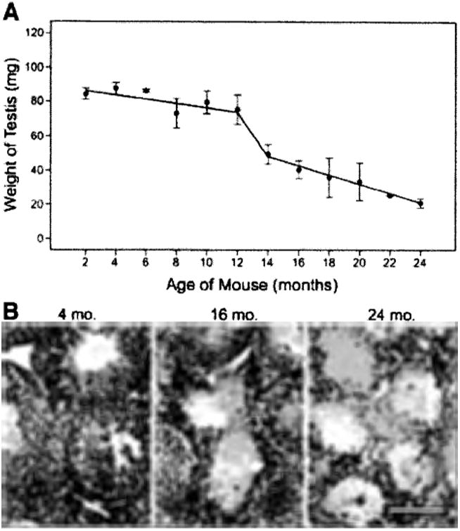
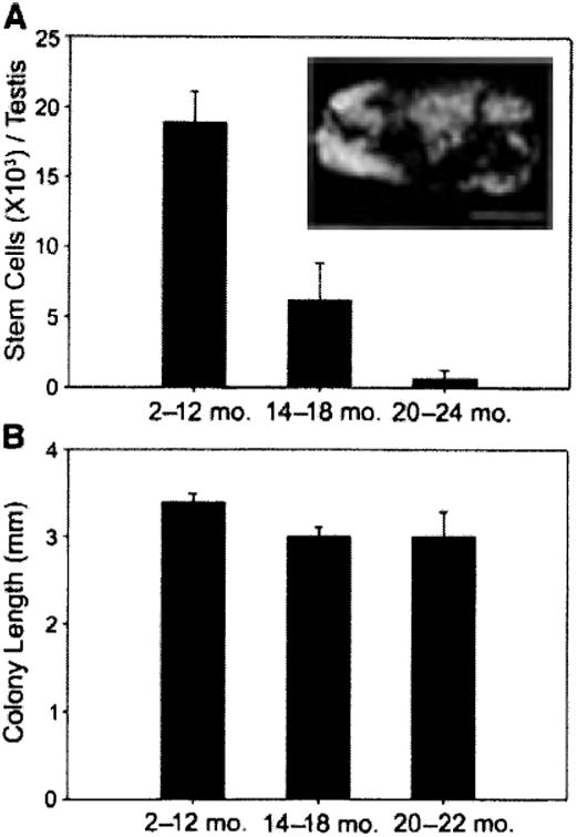
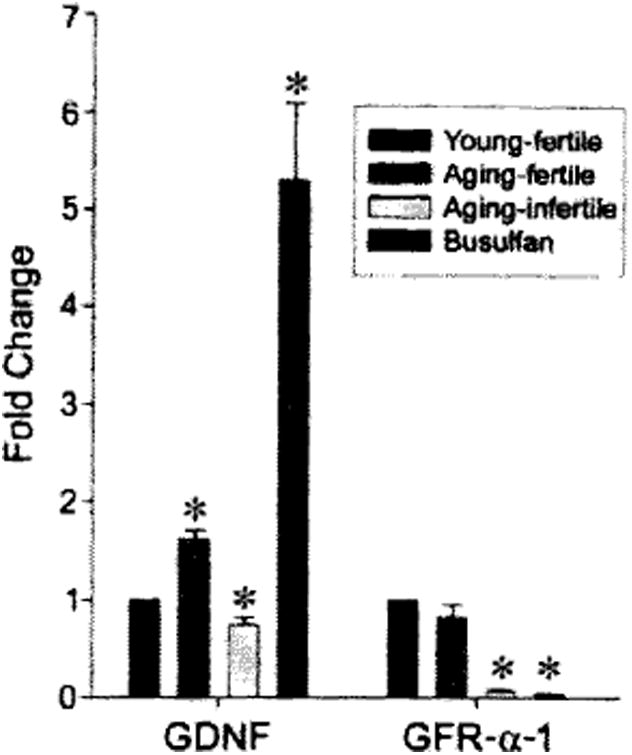
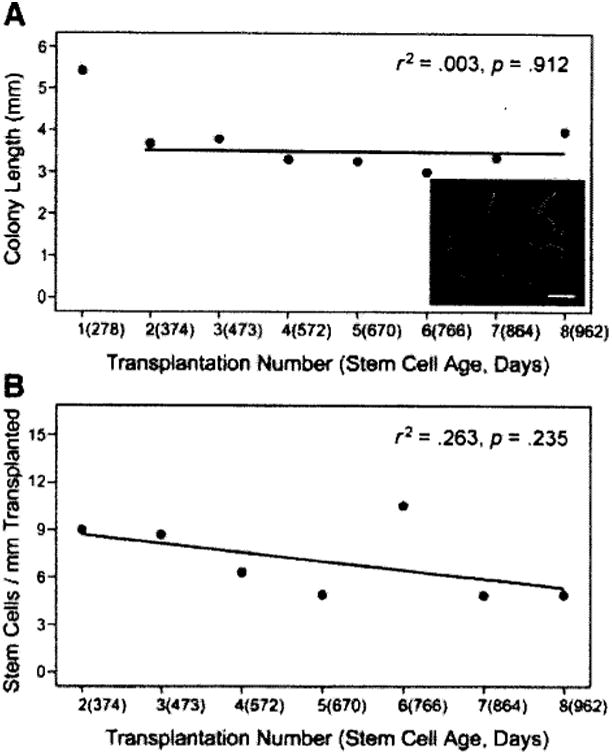
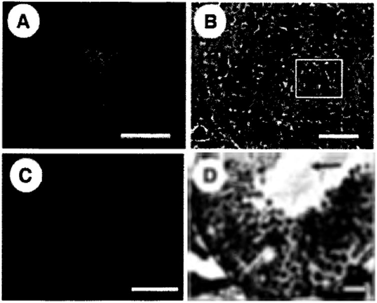
Aging is evident in most tissues and organ systems, but the mechanisms of aging are difficult to identify and poorly understood. Here, we test the hypothesis that aging results in uncorrected defects in stem cell and/or niche function, which lead to system failure. We used the spermatogonial stem cell (SSC) transplantation assay to determine the effect of aging on testis stem cell/niche function in mice. Between 12 and 24 months of age, male mice experienced a declining level of fertility associated with decreased testis weight, level of spermatogenesis, and total stem cell content. However, when stem cells were consecutively passaged at 3-month intervals to testes of young males, these stem cells continued to produce spermatogenesis for more than 3 years. Thus, SSC self-renewal continues long past the normal life span of the animal when the stem cell is continually maintained in a young niche/microenvironment. Moreover, these data suggest that infertility in old males results from deterioration of the SSC niche and failure to support an appropriate balance between stem cell self-renewal and differentiation.


Conflict of interest statement

Disclosures: The authors indicate no potential conflicts of interest.

Figures

Figure 1
Figure 1
Testis weight and morphological appearance of normal spermatogenesis decrease with age. (A): Testis weights (mg) were determined for male ROSA26 mice from 2-24 months of age. (B): Histological evaluation of testes from aging mice. Left: Young males (4 months) had complete spermatogenesis in all seminiferous tubules. Center: Testes of aging males (16 months) had tubules with spermatogenesis as well as empty tubules. Right: Spermatogenesis was absent in the seminiferous tubules of old males (24 months). Stain = hematoxylin and eosin. Scale bar = 100 μm.
Figure 2
Figure 2
Stem cell activity in mouse testes. (A, inset): Macroscopic appearance of recipient testis transplanted with donor testis cell. Individual blue tubules indicate colonies of spermatogenesis arising from donor spermatogonial stem cells (SSCs). The SSC transplantation assay was used to evaluate stem cell activity in the testes of young (2–12 months), aging (14–18 months), and old (20–24 months) mice. Donor-derived colonies of spermatogenesis are unequivocally identified in recipient seminiferous tubules because they express a reporter gene (i.e., lacZ). Stem cell number (colony number) and kinetics of colony expansion (e.g., colony length) can be evaluated. Bar = 2 mm. (A, graph): Testis stem cell content for young (18.9 ± 2.3 ×10³, n = 12 experiments, 145 recipient testes), aging (6.1 ± 2.6 ×10³, n = 7 experiments, 84 recipient testes), and old (0.7 ± 0.6 7×10³, n = 6 experiments, 68 recipient testes) ROSA26 mice (young vs, aging, p = .002; young vs. old, p = .001; aging vs. old, p = .329). Total stem cell number per donor testis = colonies per 105 cells transplanted × total cells recovered per donor testis 7 × 20 to account for transplantation efficiency [30]. (B): Colony length (mm) from donor testis cells of young (3.4 ± 0.1, n = 24 recipients, 248 colonies), aging (3.0 ± 0.1, n = 12 recipients, 120 colonies), and old (3.0 ± 0.3, n = 10 recipients, 128 colonies) males was determined in recipients by using a dissecting microscope and digital imaging system (young vs. aging, p = .111; young vs. old, p = .089; aging vs. old, p = .977). No stem cell activity was recovered from testes of 24-month-old males. Bars are mean ± SEM
Figure 3
Figure 3
Real-time PCR analysis of GDNF and GFR-α-1 expression in the testes of young, aging, and busulfan-treated mice, using TaqMan MGB assays (Applied Biosystems). Young-fertile animals (black bars) were 2–4 months old, and testes weighed 92.5 ± 2.2 mg (n = 6). Aging-fertile animals (red bars) were 15–19 months old, and testes weighed 81.1 ± 4.2 mg (n = 4). Aging-infertile animals (yellow bars) were 15–19 months old, and testes weighed 32.1 ± 0.9 mg (n = 4). Busulfan-treated animals (blue bars) were 4 months old, and testes weighed 24.7 ±1.6 mg (n = 3). Data were normalized by comparison with the transcription factor GATA4, which is constitutively expressed in Sertoli cells, and reported as fold change (mean ± SEM) relative to the young-fertile control values. Asterisk indicates significant difference from control. Abbreviations: GDNF, glial cell line-derived neurotrophic factor; GFR, glial cell-derived neurotrophic factor family receptor; PCR, polymerase chain reaction.
Figure 4
Figure 4
Serial transplantation of mouse spermatogonial stem cells. In the primary transplant, donor testis cells were obtained from 6-month-old, cryptorchid, green fluorescent protein (GFP) mice. (A): Three months after transplantation, the testes of recipient animals were evaluated for green colonies of donor spermatogenesis (inset, scale bar = 2 mm). After the initial transplant, seven more serial transplants were performed, resulting in a final donor stern cell age of 32 months (962 days). Colony number and colony length were recorded during each serial transfer. The length (mm) of each green colony was determined using an epifluorescent microscope, a GFP filter cube, and a digital imaging system. (B): Colonies per millimeter is a retrospective value that is determined based on the number of colonies produced per millimeter of green tubule observed in the previous transplant. Thus, there is no value for the first transplant.
Figure 5
Figure 5
Histological evaluation of retransplanted seminiferous tubules. Serially retransplanted (eighth transplant, 962 days) donor spermatogonial stem cells produced normal colonies (∼3, 5, and 8 months after ninth transplant, total age 1,060, 1,102, and 1,216 days) of spermatogenesis as defined by the presence of multiple germ cell layers and sperm in the seminiferous tubule lumen of busulfan-treated (A, B) or W sterile (C, D) recipient mice. (A): Whole-mount image of green colony of donor spermatogenesis (1,102 days). (B): Frozen section through a colony of donor spermatogenesis (1,060 days). Fluorescent image showing multiple layers of green donor germ cells is overlayed on the phase-contrast image of the same section. The basement membrane is clearly visible on the left side of the section (arrow), and the boxed area indicates the center of the seminiferous tubule. (C): Whole-mount image of a green colony of donor spermatogenesis in a W recipient testis (1,216 days). (D): Cross-section through colony of donor spermatogenesis in (C). Complete spermatogenesis can be seen with sperm tails in the lumen of the seminiferous tubule. Arrow indicates sperm tails in the center of the seminiferous tubule. Scale bars = 2 mm (A, C), 50 μm (B, D).

References

    1. Schofield R. The relationship between the spleen colony-forming cell and the haemopoietic stem cell. Blood Cells. 1978;4:7–25. - PubMed
    1. Rao MS, Mattson MP. Stem cells and aging: Expanding the possibilities. Mech Ageing Dev. 2001;122:713–734. - PubMed
    1. Trosko JE. Human stem cells as targets for the aging and diseases of aging processes. Med Hypotheses. 2003;60:439–447. - PubMed
    1. Van Zant G, Liang Y. The role of stem cells in aging. Exp Hematol. 2003;31:659–672. - PubMed
    1. Harrison DE. Competitive repopulation: A new assay for long-term stem cell functional capacity. Blood. 1980;55:77–81. - PubMed

Publication types

MeSH terms

Substances