Uteroglobin-related protein 1 expression suppresses allergic airway inflammation in mice
- PMID: 16456148
- PMCID: PMC2582904
- DOI: 10.1164/rccm.200503-456OC
Uteroglobin-related protein 1 expression suppresses allergic airway inflammation in mice
Abstract
Rationale: Uteroglobin-related protein (UGRP) 1, which is highly expressed in the epithelial cells of the airways, has been suggested to play a role in lung inflammation.
Objectives: The aim of study was to understand the effect of overexpressed UGRP1 on lung inflammation in a mouse model of allergic airway inflammation.
Methods: Ovalbumin-sensitized and -challenged mice, a model for allergic airway inflammation, were used in conjunction with recombinant adenovirus expressing UGRP1.
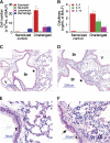
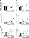
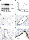
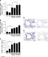
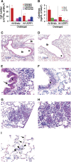
Measurements and main results: We demonstrated that intranasal administration of adeno-UGRP1 successfully delivered UGRP1 to the epithelial cells of airways and markedly reduced the number of infiltrating inflammatory cells, particularly eosinophils, in lung tissue as well as the level of proinflammatory cytokines such as interleukin (IL)-4, IL-5, and IL-13 in bronchoalveolar lavage fluids. The healed phase of inflammation was clearly seen in the peripheral areas of adeno-UGRP1-treated mouse lungs.
Conclusion: These results demonstrate that UGRP1 can suppress inflammation in the mouse model of allergic airway inflammation. Based on this result, we propose UGRP1 as a novel therapeutic candidate for treating lung inflammation such as is found in asthma.
Figures
Comment in
-
Periarterial leukocyte accumulation in allergic airway inflammation.Am J Respir Crit Care Med. 2006 Oct 1;174(7):840; author reply 840. doi: 10.1164/ajrccm.174.7.840. Am J Respir Crit Care Med. 2006. PMID: 16988165 No abstract available.
Similar articles
-
The cytokine-driven regulation of secretoglobins in normal human upper airway and their expression, particularly that of uteroglobin-related protein 1, in chronic rhinosinusitis.Respir Res. 2011 Mar 8;12(1):28. doi: 10.1186/1465-9921-12-28. Respir Res. 2011. PMID: 21385388 Free PMC article.
-
Decreased expression of uteroglobin-related protein 1 in inflamed mouse airways is mediated by IL-9.Am J Physiol Lung Cell Mol Physiol. 2004 Dec;287(6):L1193-8. doi: 10.1152/ajplung.00263.2004. Am J Physiol Lung Cell Mol Physiol. 2004. PMID: 15531759
-
Interleukin-5 reduces the expression of uteroglobin-related protein (UGRP) 1 gene in allergic airway inflammation.Immunol Lett. 2005 Feb 15;97(1):123-9. doi: 10.1016/j.imlet.2004.10.013. Immunol Lett. 2005. PMID: 15626484 Free PMC article.
-
Interleukin-10 induces uteroglobin-related protein (UGRP) 1 gene expression in lung epithelial cells through homeodomain transcription factor T/EBP/NKX2.1.J Biol Chem. 2004 Dec 24;279(52):54358-68. doi: 10.1074/jbc.M405331200. Epub 2004 Oct 13. J Biol Chem. 2004. PMID: 15485815
-
Emerging role of an immunomodulatory protein secretoglobin 3A2 in human diseases.Pharmacol Ther. 2022 Aug;236:108112. doi: 10.1016/j.pharmthera.2022.108112. Epub 2022 Jan 10. Pharmacol Ther. 2022. PMID: 35016921 Free PMC article. Review.
Cited by
-
Transgenically-expressed secretoglobin 3A2 accelerates resolution of bleomycin-induced pulmonary fibrosis in mice.BMC Pulm Med. 2015 Jul 16;15:72. doi: 10.1186/s12890-015-0065-4. BMC Pulm Med. 2015. PMID: 26178733 Free PMC article.
-
Uterus globulin associated protein 1 (UGRP1) binds podoplanin (PDPN) to promote a novel inflammation pathway during Streptococcus pneumoniae infection.Clin Transl Med. 2022 Jun;12(6):e850. doi: 10.1002/ctm2.850. Clin Transl Med. 2022. PMID: 35652821 Free PMC article.
-
The cytokine-driven regulation of secretoglobins in normal human upper airway and their expression, particularly that of uteroglobin-related protein 1, in chronic rhinosinusitis.Respir Res. 2011 Mar 8;12(1):28. doi: 10.1186/1465-9921-12-28. Respir Res. 2011. PMID: 21385388 Free PMC article.
-
Preclinical evaluation of human secretoglobin 3A2 in mouse models of lung development and fibrosis.Am J Physiol Lung Cell Mol Physiol. 2014 Jan 1;306(1):L10-22. doi: 10.1152/ajplung.00037.2013. Epub 2013 Nov 8. Am J Physiol Lung Cell Mol Physiol. 2014. PMID: 24213919 Free PMC article.
-
Plasma UGRP1 levels associate with promoter G-112A polymorphism and the severity of asthma.Allergol Int. 2008 Mar;57(1):57-64. doi: 10.2332/allergolint.O-07-493. Epub 2008 Mar 1. Allergol Int. 2008. PMID: 18089940 Free PMC article.
References
-
- Niimi T, Keck-Waggoner CL, Popescu NC, Zhou Y, Levitt RC, Kimura S. UGRP1, a uteroglobin/CCSP-related protein, is a novel lung-enriched downstream target gene for the T/EBP/NKX2.1 homeodomain transcription factor. Mol Endocrinol 2001;15:2021–2036. - PubMed
-
- Klug J, Beier HM, Bernard A, Chilton BS, Fleming TP, Lehrer RI, Miele L, Pattabiraman N, Singh G. Uteroglobin/clara 10 kDa family of proteins: nomenclature committee report. In: Mukherjee AB, Chilton BS, editors. The uteroglobin/clara cell protein family. Ann N Y Acad Sci 2000;923:348–354. - PubMed
-
- Singh G, Katyal SL. Clara cells and Clara cell 10 kD protein (CC10). Am J Respir Cell Mol Biol 1997;17:141–143. - PubMed
Publication types
MeSH terms
Substances
Grants and funding
LinkOut - more resources
Full Text Sources
Other Literature Sources
Molecular Biology Databases
Research Materials

