. 2006 Feb 22;25(4):785-97.
doi: 10.1038/sj.emboj.7600982. Epub 2006 Feb 2.

A unique set of SH3-SH3 interactions controls IB1 homodimerization

Affiliations

A unique set of SH3-SH3 interactions controls IB1 homodimerization

Ole Kristensen et al. EMBO J. .

Abstract

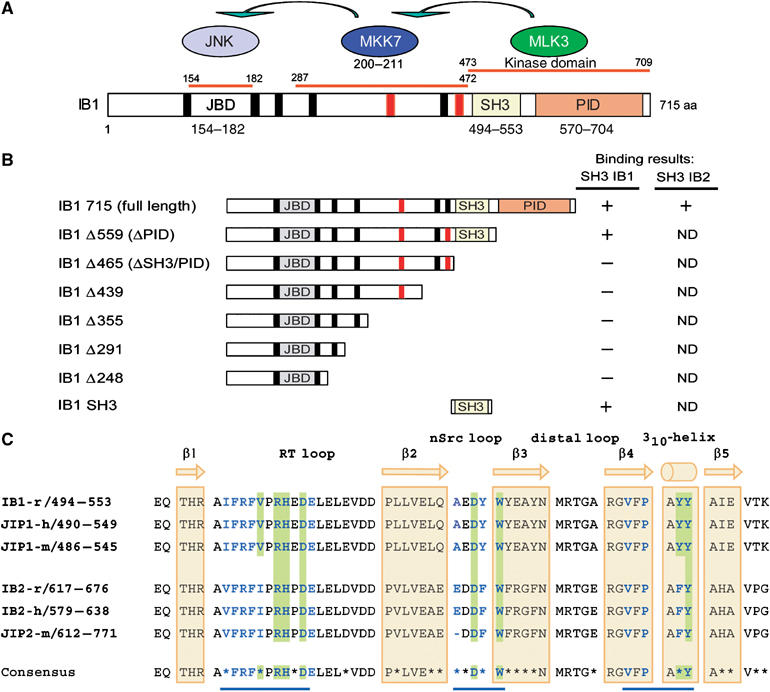
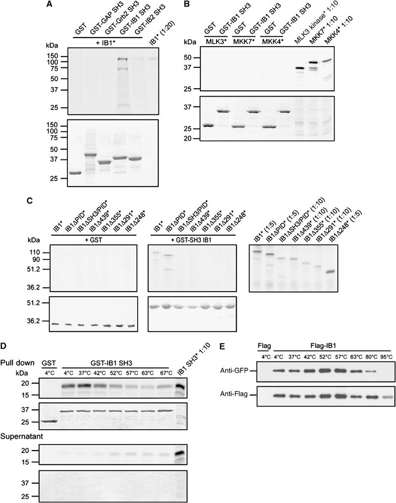
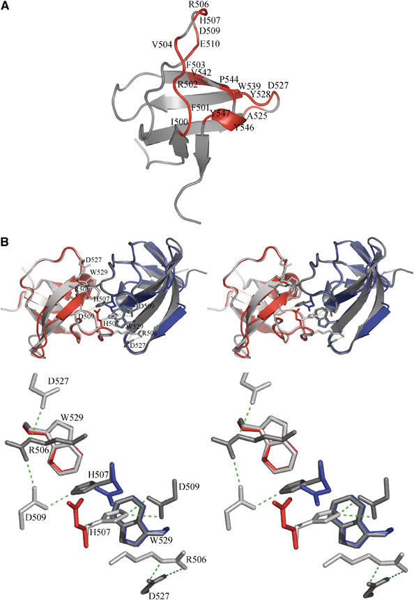
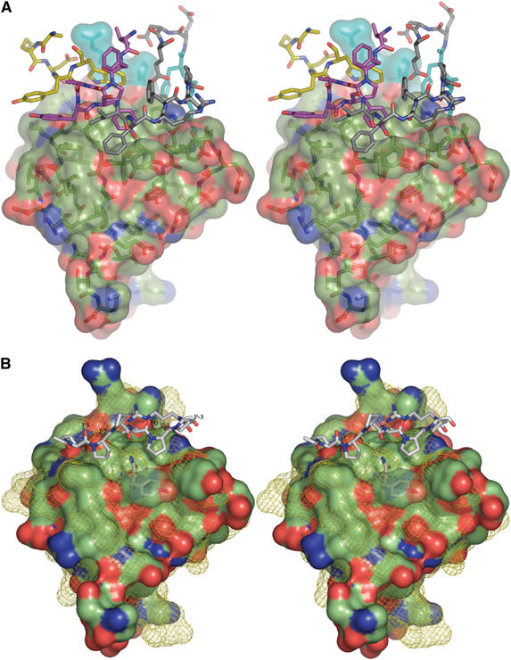
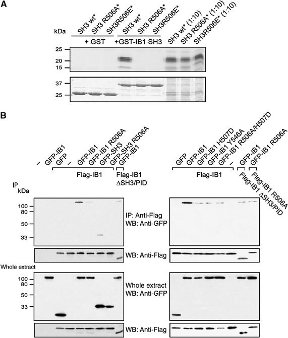
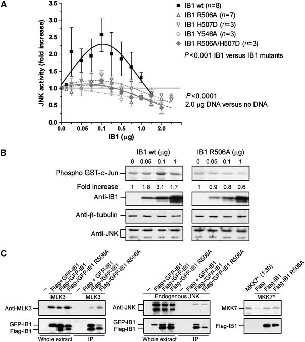
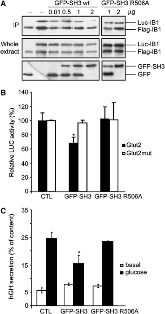
Islet-brain 1 (IB1 or JIP-1) is a scaffold protein that interacts with components of the c-Jun N-terminal kinase (JNK) signal-transduction pathway. IB1 is expressed at high levels in neurons and in pancreatic beta-cells, where it controls expression of several insulin-secretory components and secretion. IB1 has been shown to homodimerize, but neither the molecular mechanisms nor the function of dimerization have yet been characterized. Here, we show that IB1 homodimerizes through a novel and unique set of Src homology 3 (SH3)-SH3 interactions. X-ray crystallography studies show that the dimer interface covers a region usually engaged in PxxP-mediated ligand recognition, even though the IB1 SH3 domain lacks this motif. The highly stable IB1 homodimer can be significantly destabilized in vitro by three individual point mutations directed against key residues involved in dimerization. Each mutation reduces IB1-dependent basal JNK activity in 293T cells. Impaired dimerization also results in a reduction in glucose transporter type 2 expression and in glucose-dependent insulin secretion in pancreatic beta-cells. Taken together, these results indicate that IB1 homodimerization through its SH3 domain has pleiotropic effects including regulation of the insulin secretion process.


Figures

Figure 1
Figure 1
Protein interaction domains of IB1. (A) IB1 is characterized by a JBD (gray box, residues 154–182), SH3 (yellow box, residues 494–553) and a PID (orange box, residues 570–704). Residues are numbered according to the full-length rat IB1 sequence (GenBank, AF108959). The seven PxxP motifs are shown in black and red. Motifs marked in red are conserved in rat, human and mouse IB1 and IB2. MKK7 and MLK3, two of the known partners of IB1, bind to regions 287–472 and 473–709 of IB1, respectively. (B) Schematic representations of full-length and C-terminal deletion mutants of IB1. Numbering corresponds to the last amino-acid expressed in the various constructs. Binding results described in Figure 2 are summarized for all constructs. ND: not determined. (C) Sequence alignment of rat, mouse and human IB1/JIP1 and IB2/JIP2 SH3 domains. The sequence of rat IB1 (GenBank, AF108959), human JIP1 (Ensembl, ENSP00000241014), mouse JIP1 (Ensembl, ENSMUSP00000050773), rat IB2 (Ensembl, ENSRNOP00000050155), human IB2 (GenBank, AF218778) and mouse JIP2 (Ensembl, ENSMUSP00000023291) are included. The IB1 SH3 region is identical in all three species. Residues that participate to IB1 dimerization as well as those expected to do so in IB2 are shown in bold blue. Residues at the dimer interface involved in inter-protomer salt bridges or hydrogen bonds are shaded in green. Nonconserved amino acids between IB1 and IB2 are indicated with a star in the consensus sequence. The positions of the β strands 1–5 and the 310-helix are indicated in yellow.
Figure 2
Figure 2
Pull-down experiments. (A) Involvement of the SH3 domain of IB1 in dimerization. Autoradiogram of separated 35S-labeled (*) full-length IB1 pull-down products observed using the GST-fused SH3 domains of IB1, IB2, Grb2 and GAP or GST alone (upper panel). IB1 specifically recognizes the SH3 domain of IB1, but not the SH3 domains of GAP or Grb2. As expected, we observe crossbinding with the SH3 domain of IB2. (Lower panel) Sample loading control, Coomassie-stained gel. (B) Interaction of the IB1 SH3 domain with known IB1 partners. SDS–PAGE autoradiogram analysis of pull-down products obtained using GST-fused IB1 SH3 and 35S-labeled (*) MLK3, MKK7 and MKK4 (upper panel). The SH3 domain of IB1 binds neither to MLK3 and MKK7 nor to the negative control MKK4. (Lower panel) Sample loading control, Coomassie-stained gel. (C) Mapping of IB1 sequences responsible for SH3 binding. Pull-down experiments were performed with GST-IB1 SH3 and a series of 35S-labeled (*) truncated C-terminal fragments (see Figure 1B). Autoradiograms (upper panels) and Coomassie staining (lower panels) of the SDS–PAGE gels are shown. Binding is lost when the C-terminal region that includes the SH3 domain of IB1 is deleted (ΔSH3/PID). (D) Stability of the IB1 SH3 dimer. Pull-down experiments were performed with GST-IB1 SH3 and 35S-labeled (*) SH3 proteins. The washed samples were incubated at increasing temperatures (4–67°C) and analyzed by SDS–PAGE, followed by autoradiography (top panel) of the Coomassie-stained gels (bottom panel). The complex is extremely stable, with a small fraction of dimers dissociating at 52°C and above. (E) Stability of the full-length IB1 dimer. Immunoprecipitation (IP) of Flag-IB1 was performed after transfection of 293T cells with Flag or Flag-IB1 together with GFP-IB1 constructs. The washed samples were incubated at increasing temperatures and analyzed by SDS–PAGE. Cosedimented GFP-IB1 was detected by GFP immunoblotting. The amount of immunoprecipitated proteins was verified with anti-Flag antibodies. The dimer is extremely stable, with binding still detectable at 80°C.
Figure 3
Figure 3
Structure of the IB1 SH3 domain. (A) Cartoon representation of the IB1 SH3 domain (in gray). The structure is composed of five β-strands arranged into two antiparallel sheets. The first sheet is formed by strands 1 and 5 and the second sheet by the strands 2, 3 and 4. The IB1 SH3 domain forms a dimer. Residues contacting the other protomer (Table II) are colored in red and labeled. (B) Cartoon representations of the experimentally determined IB1 SH3 homodimer (gray) and of the artificial C-terminal Grb2 SH3 dimer (in red and blue) generated by superposition onto the IB1 SH3 backbone (shown in stereo). The two IB1 SH3 protomers in the dimer are in a cis arrangement and it is likely that the two PID domains are in close proximity to each other. The superimposed Grb2 dimer illustrates that in this structure two opposing glutamate residues (red and blue) are incompatible with IB1-like homodimerization (see enlargement).
Figure 4
Figure 4
Stereographic representation of the IB1 SH3 domain. (A) The solvent-accessible surface of the IB1 SH3 domain. Contact residues from the other molecule of the homodimer colored after atom type are shown as sticks. Carbon atoms from residues 500 to 510 are colored in gray, those from residues 525 to 529 are in yellow and the 542 to 547 region is shown in magenta. Residues 506 and 507 from both monomers are colored in cyan. (B) The solvent-accessible surfaces of the IB1 and SEM-5 SH3 domains. The surface of the IB1 SH3 domain is colored after atom type. The superimposed solvent-accessible surface of SEM-5 (Lim et al, 1994) is shown as a yellow mesh, along with the bound mSos-derived peptide PPPVPPR in sticks representation. The canonical PPII binding sites are labeled (Yu et al, 1994).
Figure 5
Figure 5
Mutations R506A, R506E, H507D, Y546A and R506A/H507D destabilize IB1 dimerization. (A) Pull-down experiments were performed with GST-SH3 and 35S-labeled (*) wt or mutant SH3 IB1 constructs (SH3 wt, SH3 R506A or SH3 R506E). Aliquots of 35S-labeled proteins were analyzed by SDS–PAGE and autoradiography (top panels) of the Coomassie-stained gel (bottom panels). (B) Flag-tagged IB1 was immunoprecipitated from extracts of 293T cells co-transfected with Flag-IB1 constructs (Flag-IB1 or Flag-IB1 ΔSH3/PID) and full-length wt or mutant GFP-IB1 constructs (GFP-IB1 wt, GFP-IB1 R506A, GFP-IB1 H507D, GFP-IB1 Y546A or GFP-IB1 R506A/H507D). Similar experiments were performed from cells co-transfected with Flag-IB1 and wt or mutant GFP-SH3 fusions (GFP-SH3, GFP-SH3 R506A). Co-immunoprecipitated proteins and immunoprecipitation efficiency were assessed with anti-GFP and anti-Flag antibodies, respectively. Transfection efficiencies were verified by anti-Flag and anti-GFP antibodies. IP: immunoprecipitation; WB: western blot.
Figure 6
Figure 6
IB1 dimerization increases JNK basal activity. (A) The 293T cells were transiently transfected with increasing levels of wt (IB1 wt) or mutant IB1 (IB1 R506A, IB1 H507D, IB1 Y546A or IB1 R506A/H507D). JNK activity was measured in cell extracts using GST-c-Jun as substrate. The maximal fold increase of basal JNK activity is reached in cells transfected with 0.1 μg of wt IB1 DNA. (B) Transfection efficiencies and protein quantification were verified by Western blotting with anti-IB1, anti-β-tubulin and anti-JNK antibodies as indicated. (C) Relative binding of JNK, MKK7 and MLK3 to wt or mutant IB1. Flag-tagged IB1 was immunoprecipitated from extracts of 293T cells co-transfected with Flag- and GFP-IB1 constructs (Flag-IB1 wt, Flag-IB1 R506A or Flag and GFP-IB1 wt or GFP-IB1 R506A) in the presence or absence of MLK3-pCMVsport6 construct. Co-immunoprecipitated MLK3 or endogenous JNK was assessed with anti-MLK3 and anti-JNK antibodies, respectively. Pull-down experiments were performed with immunoprecipitated Flag, wt or mutant Flag-IB1 and 35S-labeled (*) MKK7. Binding of MKK7 was detected by autoradiography.
Figure 7
Figure 7
Functional role of the IB1 dimer. (A) Overexpression of the IB1 SH3 domain results in destabilization of the IB1 dimer. INS-1E cells were co-transfected with Flag-IB1, Luc-IB1 and increasing amount of GFP-SH3 wt and mutant constructs. Flag-tagged IB1 was immunoprecipitated from INS-1E extracts. Co-immunoprecipitated Luc-IB1 and immunoprecipitation efficiency were assessed with anti-IB1 antibodies. Transfection efficiencies were verified by anti-IB1 and anti-GFP antibodies. IP: immunoprecipitation. (B) INS-1E cells were transiently co-transfected with a luciferase-encoding plasmid driven by the Glut2 promoter (Glut2) or by a mutant form (Glut2mut), with an empty plasmid (control) or wt or mutant GFP-tagged SH3 expression vectors (GFP-SH3 wt, GFP-SH3 R506A). The relative luciferase activities are reported. Overexpression of wt SH3 leads to a significant decrease in the expression of GLUT2, whereas the mutated domain did not have any effect. (C) Dimerization of IB1 is required for glucose-induced insulin secretion. INS-1E cells were transiently co-transfected with an hGH-encoding plasmid and with an empty plasmid (control) or a wt or mutant GFP-tagged SH3 expression vectors (GFP-SH3 wt, GFP-SH3 R506A). Cells were later incubated under either basal or stimulatory conditions. The total amount of hGH present in the cells and the fraction released in the medium were measured by ELISA. Overexpression of wt SH3 leads to a significant decrease in stimulated secretion, whereas the mutated domain had no effect.

References

    1. Bonny C, Nicod P, Waeber G (1998) IB1, a JIP-1-related nuclear protein present in insulin-secreting cells. J Biol Chem 273: 1843–1846 - PubMed
    1. Bonny C, Oberson A, Negri S, Sauser C, Schorderet DF (2001) Cell-permeable peptide inhibitors of JNK: novel blockers of beta-cell death. Diabetes 50: 77–82 - PubMed
    1. Bonny C, Oberson A, Steinmann M, Schorderet DF, Nicod P, Waeber G (2000) IB1 reduces cytokine-induced apoptosis of insulin-secreting cells. J Biol Chem 275: 16466–16472 - PubMed
    1. Borsello T, Clarke PG, Hirt L, Vercelli A, Repici M, Schorderet DF, Bogousslavsky J, Bonny C (2003) A peptide inhibitor of c-Jun N-terminal kinase protects against excitotoxicity and cerebral ischemia. Nat Med 9: 1180–1186 - PubMed
    1. Brasher BB, Roumiantsev S, Van Etten RA (2001) Mutational analysis of the regulatory function of the c-Abl Src homology 3 domain. Oncogene 20: 7744–7752 - PubMed

Publication types

MeSH terms