. 2006 Feb 22;25(4):727-38.
doi: 10.1038/sj.emboj.7600980. Epub 2006 Feb 2.

B56-containing PP2A dephosphorylate ERK and their activity is controlled by the early gene IEX-1 and ERK

Affiliations

B56-containing PP2A dephosphorylate ERK and their activity is controlled by the early gene IEX-1 and ERK

Claire Letourneux et al. EMBO J. .

Abstract

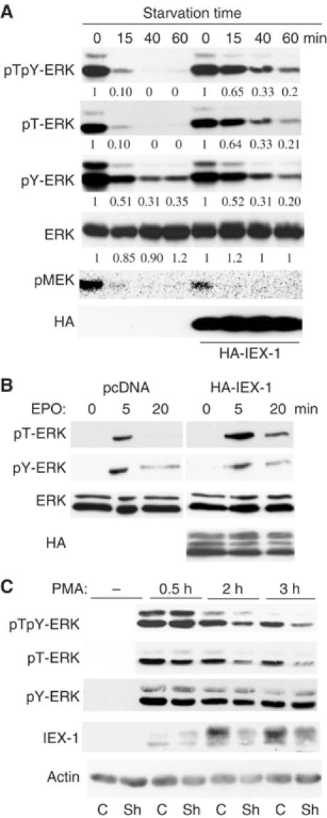
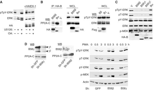
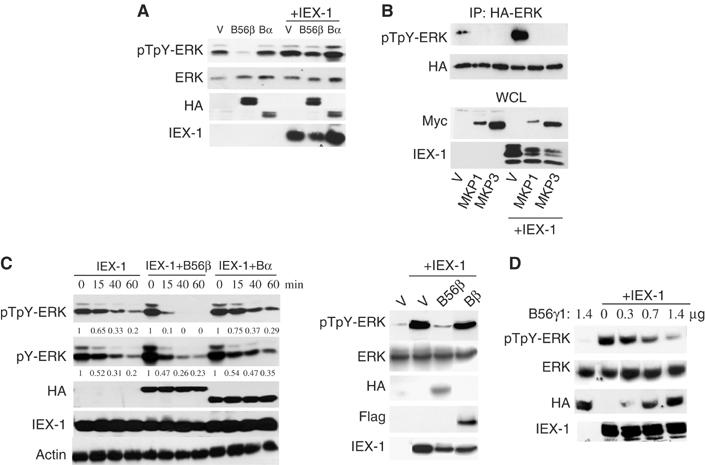
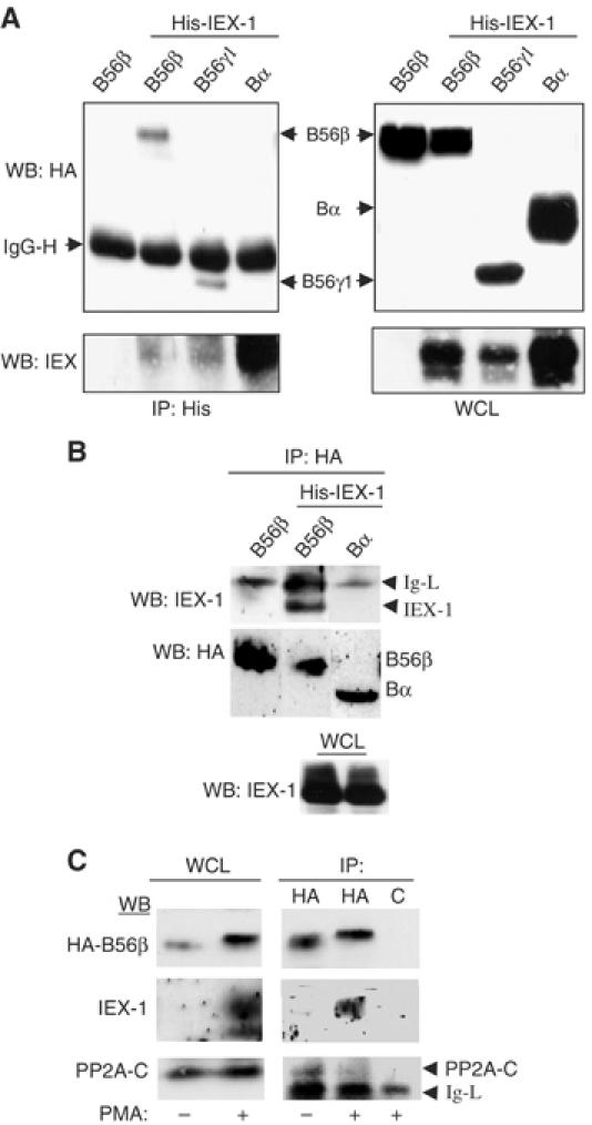
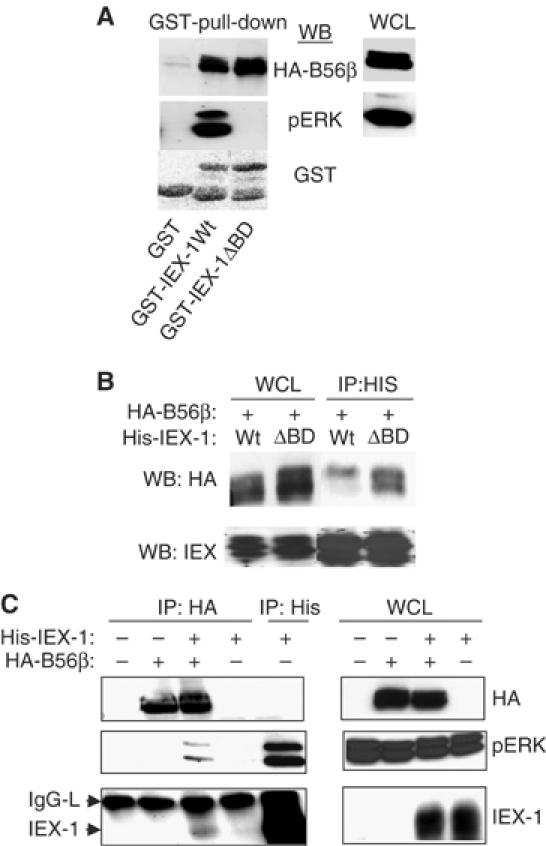
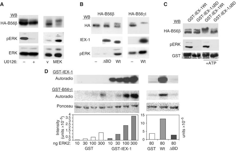
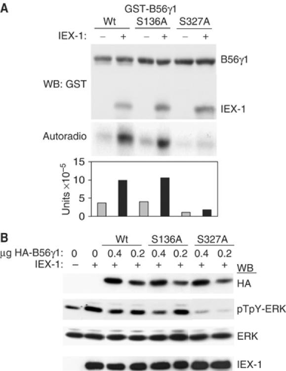
The protein phosphatase 2A (PP2A) acts on several kinases in the extracellular signal-regulated kinase (ERK) signaling pathway but whether a specific holoenzyme dephosphorylates ERK and whether this activity is controlled during mitogenic stimulation is unknown. By using both RNA interference and overexpression of PP2A B regulatory subunits, we show that B56, but not B, family members of PP2A increase ERK dephosphorylation, without affecting its activation by MEK. Induction of the early gene product and ERK substrate IEX-1 (ier3) by growth factors leads to opposite effects and reverses B56-PP2A-mediated ERK dephosphorylation. IEX-1 binds to B56 subunits and pERK independently, enhances B56 phosphorylation by ERK at a conserved Ser/Pro site in this complex and triggers dissociation from the catalytic subunit. This is the first demonstration of the involvement of B56-containing PP2A in ERK dephosphorylation and of a B56-specific cellular protein inhibitor regulating its activity in an ERK-dependent fashion. In addition, our results raise a new paradigm in ERK signaling in which ERK associated to a substrate can transphosphorylate nearby proteins.


Figures

Figure 1
Figure 1
IEX-1 inhibits ERK dephosphorylation on threonine. (A, B) IEX-1 expression extends the pT-ERK signal. CHO-EpoR cells were transfected with either empty vector or HA-IEX-1. The cells were either stimulated with FCS for 5 min and then starved for various times (A) or stimulated for various times with 10 U/ml Epo (B). Quantification of the pERK levels in cells expressing IEX-1 or control, normalized to ERK levels, is expressed relative to the levels of phosphorylation at their zero time point. (C) IEX-1 downregulation reduces pT-ERK signal duration. HeLa cells expressing shRNA for IEX-1 (Sh) or GFP (C) were starved of FCS overnight and stimulated with PMA (20 ng/ml). ERK phosphorylation and IEX-1 expression were examined at various times following stimulation.
Figure 2
Figure 2
B56 subunits containing PP2A dephosphorylate ERK. (A) The PP2A inhibitor OA prevents ERK dephosphorylation. CHO cells were treated for 30 min at 37°C with 10 μM U0126 or vehicle alone, in the presence or absence of 0.1 μM OA and analyzed for ERK phosphorylation. (B, C) The B56- but not the B-family of PP2A dephosphorylates ERK. NIH-3T3 (B) or CHO (C) cells were transfected with empty vector (V) or the various PP2A-B subunits (4 × HA-B56β, HA-Bα, Flag-Bβ, Flag-Bγ, HA-B56γ). Anti-HA immunoprecipitates were blotted with anti-PP2A-C (B, left panel). ERK phosphorylation was analyzed in the whole-cell lysates (WCL). (D) Downregulation of B56-PP2A leads to increased ERK phosphorylation. Left panel: Expression of B56γ and B56β subunits in HeLa cells expressing the corresponding shRNA. Right panel: Cells expressing B56γ, B56β or control (GFP) shRNAs were starved of FCS overnight and stimulated with 10 ng/ml PMA for various times.
Figure 3
Figure 3
Competition between IEX-1 and B56-containing PP2A in ERK signaling. (A, B) IEX-1 reverses B56β- but not MKP-mediated ERK dephosphorylation. CHO cells were transfected with 1 μg of the various HA-B subunits (A) or with 0.7 μg HA-ERK and 0.7 μg Myc-MKP1/3 (B) in the presence of 1 μg of pcDNA (V) or His-IEX-1. ERK phosphorylation was detected 24 h following transfection in WCL (A) or anti-HA precipitates (B). (C, D) B56 but not B subunits compete with IEX-1 effect on ERK activation. (C) CHO-EpoR cells were transfected with 0.5 μg His-IEX-1 together with 0.5 μg of HA- or Flag-tagged B subunits. The cells were stimulated and starved as depicted in legend to Figure 1A. Quantification of pERK levels is shown relative to the levels of phosphorylation at zero time point; after normalization to actin levels. (D) ERK activation was assessed in CHO cells transfected with 0.4 μg of IEX-1 and the indicated amounts of HA-B56γ1 or empty vector.
Figure 4
Figure 4
IEX-1 interacts with B56 but not with B subunits. (A, B) CHO cells were transfected with His-IEX-1 together with the indicated HA-B subunits. After 24 h, the lysates were immunoprecipitated with anti-His (A) or anti-HA (B) antibodies. (C) HeLa cells stably expressing HA-B56β were treated or not for 2 h with PMA to induce IEX-1 expression and the lysates were precipitated with either anti-HA or control (c) antibodies. The presence of B proteins and IEX-1 in the immunoprecipitates and WCL was detected by Western blotting.
Figure 5
Figure 5
IEX-1 forms a ternary complex with B56 and perk. (A) Lysates from CHO cells transfected with HA-B56β were reacted with Sepharose-bound-GST, GST-IEX-1-Wt or GST-IEX-1-ΔBD for 1 h at 4°C and washed. The presence of pERK and B56β was analyzed. (B, C) CHO cells were transfected with His-IEX-1 (Wt or ΔBD) HA-B56β subunit alone or together. The indicated antibodies were used to blot anti-His (B, C) and anti-HA (C) immunoprecipitates and WCL.
Figure 6
Figure 6
IEX-1 increases the phosphorylation of B56 subunits by ERK. (A, B) ERK-dependent phosphorylation of B56 subunits. CHO cells were transfected with HA-B56β or B56γ1, alone or together with either constitutively active MEK (A) or IEX-1 (B). The shift in B56 mobility and IEX-1 and pERK expressions were analyzed on 7 and 10% acrylamide SDS gels, respectively. (A) U0126 (10 μM) was added 40 min before cell lysis. (C) IEX-1-associated ERK induces B56β mobility shift. Lysates from CHO cells transfected with HA-B56β were reacted with Sepharose-bound GST-IEX-1-Wt or GST-IEX-1-ΔBD. After six washes, the beads were incubated at 30°C for 20 min in kinase buffer containing or not ATP before Western blotting with the indicated antibodies. (D) IEX-1 increases ERK activity towards the B56γ1 subunit in vitro. Then, 1 μg GST-B56γ1 was subjected to in vitro kinase assays with the indicated amounts of recombinant ERK2, in the presence of 1 μg of either GST, GST-IEX-1-Wt or GST-IEX-1-ΔBD. Autoradiographies of GST-B56 and GST-IEX-1 are shown. The plots represent quantification of the radioactivity incorporated into GST-B56γ. Ponceau staining shows equal loading of GST-B56γ1.
Figure 7
Figure 7
ERK-mediated phosphorylation of B56γ1 at Ser327 leads to inhibition of its activity towards ERK. (A) Identification of the ERK phosphorylation site in B56γ1. GST-B56γ1 Wt, S136A or S327A were subjected to in vitro kinase assays with 100 ng ERK, in the presence or absence of GST-IEX-1, as in legend to Figure 6D. The plot shows the radioactivity incorporated in B56γ1, normalized to the levels of the GST detected in the anti-GST blot. (B) Effect of B56γ1 mutants on ERK activation. CHO cells were tansfected with 0.4 μg of IEX-1 together with various amounts of the indicated B56γ1 forms and assayed for ERK phosphorylation.
Figure 8
Figure 8
IEX-1 induces ERK-dependent dissociation of B56/A/C trimers. (A–C) IEX-1 displaces B56 from the PP2A-C subunit. CHO cells were transfected with 1 μg of HA-B56β (A, B) or HA-B56γ1 (C) and either 2 μg of pcDNA, His-IEX-1-Wt or His-IEX-1-ΔBD. The anti-HA (A, C) or anti-PP2A-C (B) precipitates and WCL were blotted with the indicated antibodies. (D) ERK-mediated phosphorylation of B56γ1 decreases its interaction with PP2A-A/C. Sepharose-bound GST-B56γ1 was phosphorylated (+ERK2-PP) or not in vitro with ERK2, and incubated for 2 h at 4°C with CHO cell lysates. The presence of endogenous PP2A-A and C subunits in the precipitates was assessed. Autoradiography indicates the phosphorylation of GST-B56γ1. (E, F) IEX-1 interacts with the PP2A-C subunit when ERK is not activated. CHO cells transfected with HA-B56β (D) or HA-IEX-1 (E) were starved or not (FCS) of serum for 4 h. The lysates were either precipitated with Sepharose bound-GST fusion proteins (D) or anti-HA antibodies.
Figure 9
Figure 9
A model for the control of ERK signal by PP2A and IEX-1. (A) Early time points of growth factor stimulation. The high ERK activation by upstream kinases overcomes the activity of B56-PP2A and the signal proceeds. (B, C) Late time points of growth factor stimulation. Upstream activation of ERK is low and B56-PP2A activity overcomes that of ERK, leading signal arrest. In cells where IEX-1 is induced (C), inhibition of B56-PP2A leads to sustained ERK signal (see text for details).

References

    1. Adams DG, Coffee RL Jr, Zhang H, Pelech S, Strack S, Wadzinski BE (2005) Positive regulation of Raf1-MEK1/2-ERK1/2 signaling by protein serine/threonine phosphatase 2A holoenzymes. J Biol Chem 280: 42644–42654 - PubMed
    1. Alessi DR, Gomez N, Moorhead G, Lewis T, Keyse SM, Cohen P (1995) Inactivation of p42 MAP kinase by protein phosphatase 2A and a protein tyrosine phosphatase, but not CL100, in various cell lines. Curr Biol 5: 283–295 - PubMed
    1. Brondello JM, Pouyssegur J, McKenzie FR (1999) Reduced MAP kinase phosphatase-1 degradation after p42/p44MAPK-dependent phosphorylation. Science 286: 2514–2517 - PubMed
    1. Camps M, Nichols A, Gillieron C, Antonsson B, Muda M, Chabert C, Boschert U, Arkinstall S (1998) Catalytic activation of the phosphatase MKP-3 by ERK2 mitogen-activated protein kinase. Science 280: 1262–1265 - PubMed
    1. Chen J, St-Germain JR, Li Q (2005) B56 regulatory subunit of protein phosphatase 2A mediates valproic acid-induced p300 degradation. Mol Cell Biol 25: 525–532 - PMC - PubMed

Publication types

MeSH terms

Associated data