. 2006 Feb 22;25(4):701-12.
doi: 10.1038/sj.emboj.7600974. Epub 2006 Feb 2.

Molecular analysis of receptor protein tyrosine phosphatase mu-mediated cell adhesion

Affiliations

Molecular analysis of receptor protein tyrosine phosphatase mu-mediated cell adhesion

Alexandru Radu Aricescu et al. EMBO J. .

Abstract

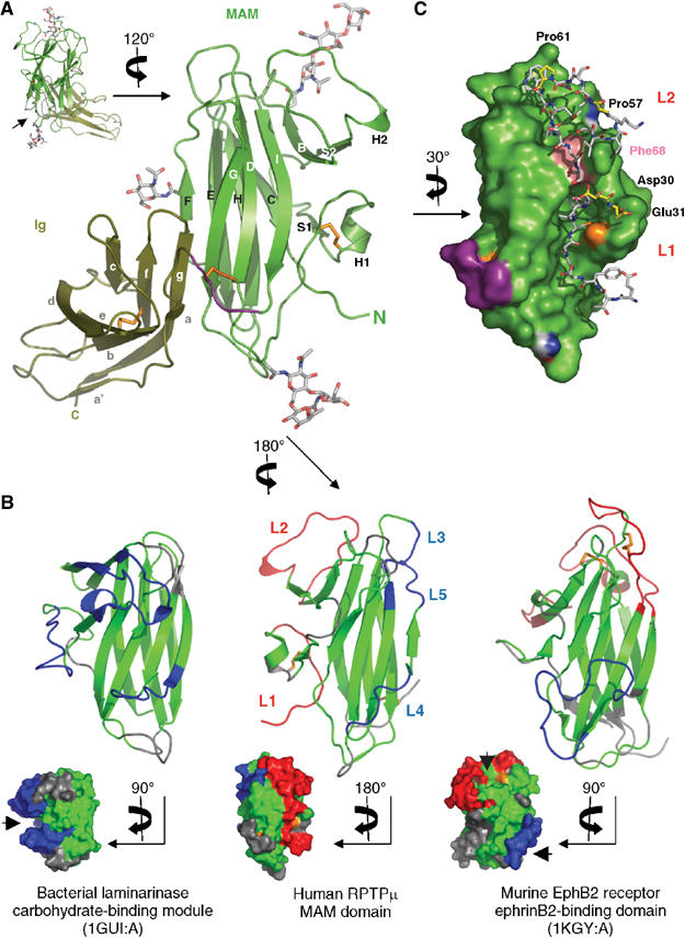
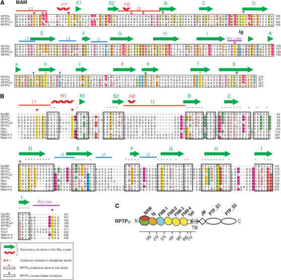
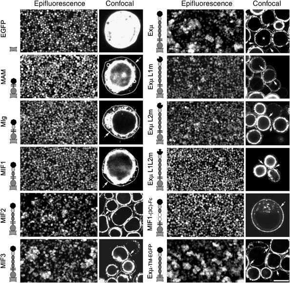
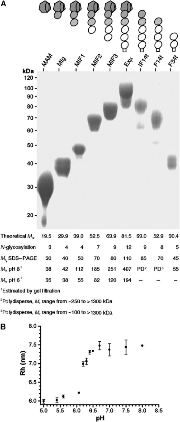
Type IIB receptor protein tyrosine phosphatases (RPTPs) are bi-functional cell surface molecules. Their ectodomains mediate stable, homophilic, cell-adhesive interactions, whereas the intracellular catalytic regions can modulate the phosphorylation state of cadherin/catenin complexes. We describe a systematic investigation of the cell-adhesive properties of the extracellular region of RPTPmu, a prototypical type IIB RPTP. The crystal structure of a construct comprising its N-terminal MAM (meprin/A5/mu) and Ig domains was determined at 2.7 A resolution; this assigns the MAM fold to the jelly-roll family and reveals extensive interactions between the two domains, which form a rigid structural unit. Structure-based site-directed mutagenesis, serial domain deletions and cell-adhesion assays allowed us to identify the four N-terminal domains (MAM, Ig, fibronectin type III (FNIII)-1 and FNIII-2) as a minimal functional unit. Biophysical characterization revealed at least two independent types of homophilic interaction which, taken together, suggest that there is the potential for formation of a complex and possibly ordered array of receptor molecules at cell contact sites.


Figures

Figure 1
Figure 1
The MIg crystal structure and analysis of the MAM domain. (A) Ribbon diagram of MIg. The MAM-Ig linker in the MAM domain is highlighted in purple. Disulphide bonds (orange) and the N-glycosylation sites (CPK) are presented as stick models. The N- and C-termini are labelled. Inset shows the L-shape of the molecule. Arrowhead indicates the largest crystal contact site. (B) Comparisons of the MAM domain to two closely related β-sandwich structures. Ribbon diagrams are shown for comparisons of secondary structures and molecular surfaces are shown to display surface features of binding sites (marked by arrowheads). Structurally equivalent regions (inter-Cα distances <3.0 Å) are shown in green and structurally distinct regions are highlighted (blue in front of the β-sandwich and red at the back). In 1GUI:A, the blue loops demarcate the carbohydrate-binding groove. In 1KGY:A, the red loops surround the hydrophobic ephrinB2-binding pocket, which constitutes the primary dimerization site; the blue loop forms the second ephrinB2-binding site. Regions that are not superposable are coloured in grey. Disulphide bonds are shown as orange sticks. All three structures are shown from the same view upon superimposition on MAM. (C) Structural details of the MAM L1 and L2 loops. The L1 and L2 loops are depicted in stick representation whereas the remainder of the MAM domain is shown as a solid surface. Residues selected for mutagenesis studies are highlighted with yellow carbon atoms. Phe68 (shown in pink) corresponds to the F74S cancer-linked mutation in RPTPρ. As in panel A, linker residues are coloured in purple and cysteines in orange. Asparagine residues providing sites for N-linked glycosylation are distinguished by standard atom colouring (carbon: white, nitrogen: blue, oxygen: red). This figure was produced using Pymol (http://pymol.sourceforge.net/).
Figure 2
Figure 2
Sequence alignments and secondary structure elements of the MAM and Ig domains. (A) Sequence alignment of the type IIB RPTPs MAM and Ig domains. Residue numbering begins with the signal sequence (not shown). Residues with more than three occurrences of sequence identity are highlighted and coloured by type. Conserved cysteines are marked by the symbol ‘▾' with disulphide pairs in the same colour and secondary structure elements are shown above the sequence. Special regions (denoted by thick lines) discussed in this report are also highlighted. Mutations in the MAM-specific loops (in red) and positions corresponding to RPTPρ cancer-linked mutations discussed in the text (in black) are labeled. (B) Sequence alignment of the MAM domains from representative human proteins. Type IIB RPTPs, neuropilins (Nrp) and meprins are shown. Regions of the ligand-binding domain of Ephrin B2 receptor (EphB2, 1KGY:A) that are structurally equivalent to the RPTPμ MAM domain are also aligned (framed). The symbols ‘*' and ‘^' denote invariant and highly conserved residues among the human Ephrin receptors. A Phe residue (in blue frame) can be considered invariantly conserved between the two folds. The two disulphide pairs and the secondary structures of the RPTPμ MAM are also displayed. (C) Schematic representation of the domain structure of full-length RPTPμ. Keys: TM/JM=transmembrane/juxtamembrane region, PTP_D1/D2=first/second phosphatase domain.
Figure 3
Figure 3
Cell-adhesion assays. Insect Sf9 cells expressing transmembrane RPTPμ EGFP-fusion constructs were observed by fluorescence microscopy. All RPTPμ fusion constructs (except for Exμ-TM-EGFP) have JM-D1-EGFP in the intracellular region, and are expressed at the plasma membrane (indicated by white arrows); whereas EGFP alone (control) is expressed uniformly in the cytosol. The bright intracellular signal indicates overexpression of the constructs in the endoplasmic reticulum and the Golgi apparatus. Note the localization of MIF2, MIF3, Exμ and its mutants (L1m, L2m) at the cell–cell contact regions (also see Figure 5B and C). IF14t and F14t fusion constructs (not shown) did not induce cell aggregation. The Exμ-TM-EGFP construct contains only the first 10 intracellular residues of RPTPμ. Scale bar: 250 μm for the epifluorescence images; 20 μm (for constructs that form cell aggregates) and 10 μm (others) for the confocal images.
Figure 4
Figure 4
Oligomeric states of RPTPμ ectodomain constructs. (A) Schematic representation of the deletion constructs and Coomassie-stained gel showing the purified soluble constructs, and estimated Mr. The indicated number of N-glycosylation sites is based on computer analysis. (B) Evaluation of pH-dependent homodimerization of non-His6-tagged Exμ by dynamic light scattering. The averaged hydrodynamic radius (Rh) is plotted against solution pH. Error bars represent standard errors of triplicate measurements.
Figure 5
Figure 5
Hypothetical schemes of Exμ adhesive interactions. (A) Possible arrangements of Exμ homophilic interactions that are consistent with the presence of two adhesion sites in the MIF1 region (one pH-dependent and one involving the L1/L2 loops). The FNIII-2 is required for efficient cell adhesion—possibly by facilitating the extension of an adhesive zipper. (B) 3D view of insect cells expressing the Exμ-TM-D1-EGFP construct. Note the extended planarity of the plasma membrane at the intercellular contact sites (arrowheads) that may be indicative of a 2D-adhesive array. The slab of cell layer (3.6 μm in depth) amidst a cell aggregate was imaged 0.2 μm apart by fluorescence confocal microscopy. This projection view is tilted 15° with respect to the 2D, single image shown in Figure 3 (‘*' denotes the same cell in the two images). (C) Cartoon outlining the cellular structures observed in the (B) image: plasma membrane (blue), endoplasmic reticulum and Golgi apparatus (green).

Similar articles

Cited by

References

    1. Andreeva A, Howorth D, Brenner SE, Hubbard TJ, Chothia C, Murzin AG (2004) SCOP database in 2004: refinements integrate structure and sequence family data. Nucleic Acids Res 32 (Database issue): D226–D229 - PMC - PubMed
    1. Aricescu AR, Fulga TA, Cismasiu V, Goody RS, Szedlacsek SE (2001) Intramolecular interactions in protein tyrosine phosphatase RPTPmu: kinetic evidence. Biochem Biophys Res Commun 280: 319–327 - PubMed
    1. Beckmann G, Bork P (1993) An adhesive domain detected in functionally diverse receptors. Trends Biochem Sci 18: 40–41 - PubMed
    1. Beltran PJ, Bixby JL (2003) Receptor protein tyrosine phosphatases as mediators of cellular adhesion. Front Biosci 8: d87–d99 - PubMed
    1. Bianchi C, Sellke FW, Del Vecchio RL, Tonks NK, Neel BG (1999) Receptor-type protein-tyrosine phosphatase mu is expressed in specific vascular endothelial beds in vivo. Exp Cell Res 248: 329–338 - PubMed

Publication types

MeSH terms

Substances