. 2006 Feb 22;25(4):713-26.
doi: 10.1038/sj.emboj.7600973. Epub 2006 Feb 2.

MAPKAPK-2-mediated LIM-kinase activation is critical for VEGF-induced actin remodeling and cell migration

Affiliations

MAPKAPK-2-mediated LIM-kinase activation is critical for VEGF-induced actin remodeling and cell migration

Miho Kobayashi et al. EMBO J. .

Abstract

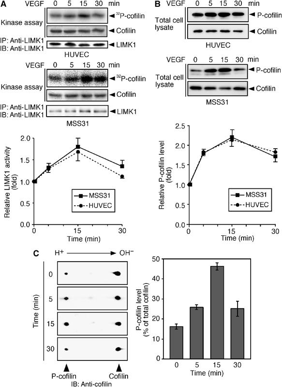
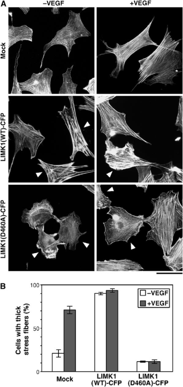
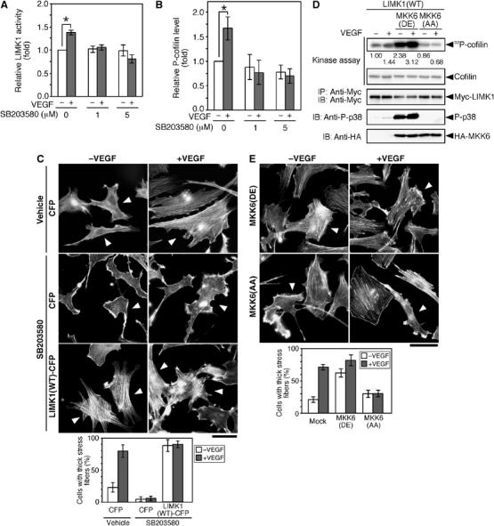
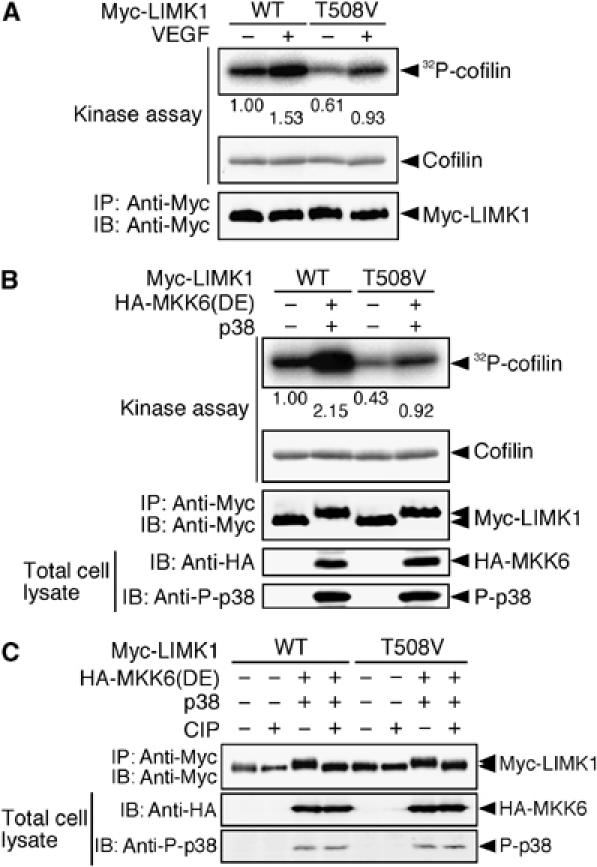
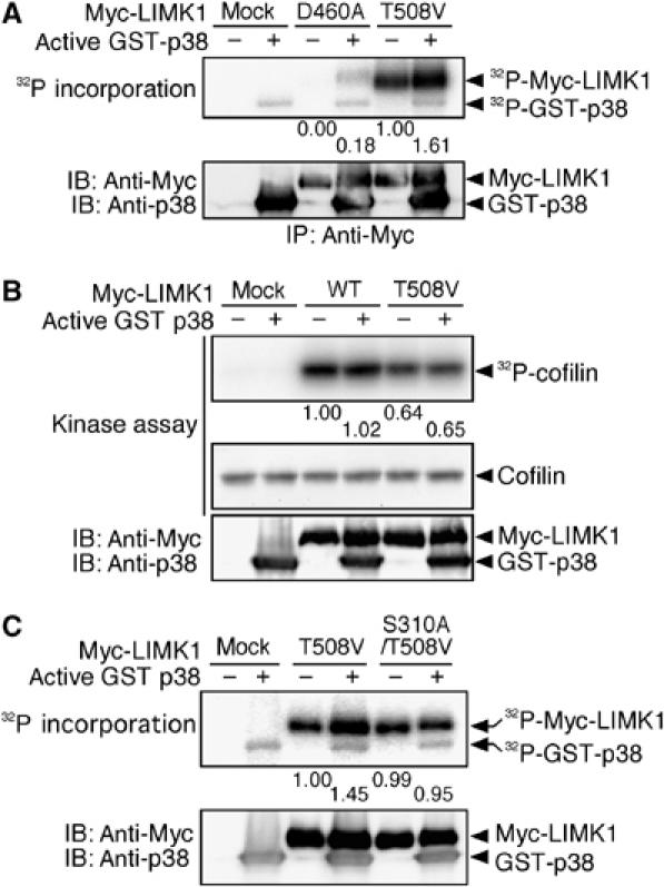
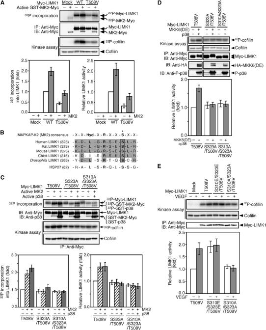
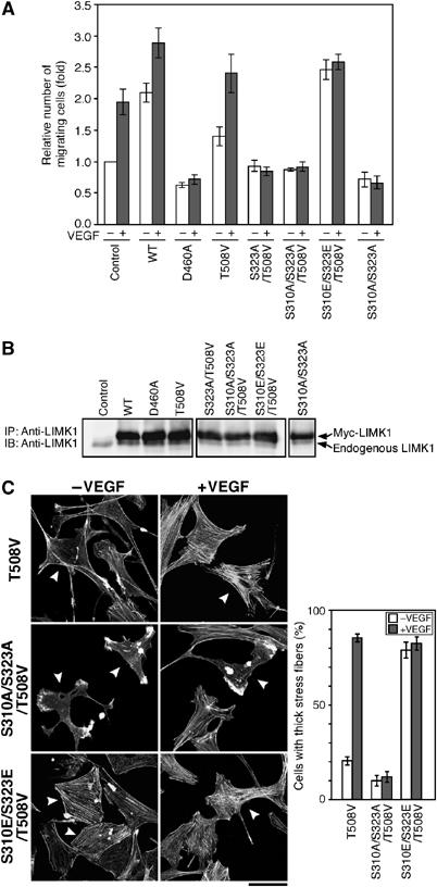
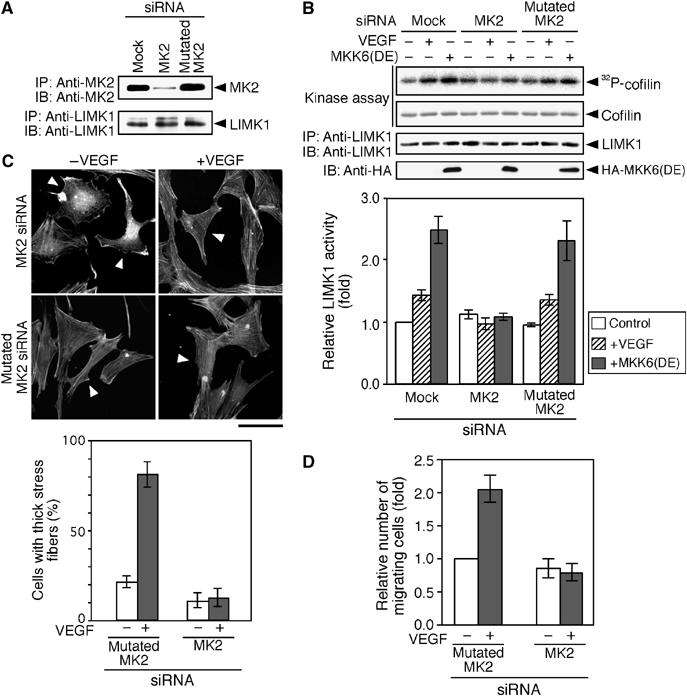
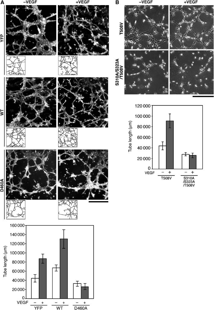
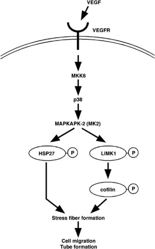
Vascular endothelial growth factor-A (VEGF-A) induces actin reorganization and migration of endothelial cells through a p38 mitogen-activated protein kinase (MAPK) pathway. LIM-kinase 1 (LIMK1) induces actin remodeling by phosphorylating and inactivating cofilin, an actin-depolymerizing factor. In this study, we demonstrate that activation of LIMK1 by MAPKAPK-2 (MK2; a downstream kinase of p38 MAPK) represents a novel signaling pathway in VEGF-A-induced cell migration. VEGF-A induced LIMK1 activation and cofilin phosphorylation, and this was inhibited by the p38 MAPK inhibitor SB203580. Although p38 phosphorylated LIMK1 at Ser-310, it failed to activate LIMK1 directly; however, MK2 activated LIMK1 by phosphorylation at Ser-323. Expression of a Ser-323-non-phosphorylatable mutant of LIMK1 suppressed VEGF-A-induced stress fiber formation and cell migration; however, expression of a Ser-323-phosphorylation-mimic mutant enhanced these processes. Knockdown of MK2 by siRNA suppressed VEGF-A-induced LIMK1 activation, stress fiber formation, and cell migration. Expression of kinase-dead LIMK1 suppressed VEGF-A-induced tubule formation. These findings suggest that MK2-mediated LIMK1 phosphorylation/activation plays an essential role in VEGF-A-induced actin reorganization, migration, and tubule formation of endothelial cells.


Figures

Figure 1
Figure 1
VEGF induces LIMK1 activation and cofilin phosphorylation. (A) LIMK1 activation. HUVEC and MSS31 cells were stimulated with VEGF. At the indicated time, cells were lysed and LIMK1 was immunoprecipitated (IP) with anti-LIMK1, and subjected to an in vitro kinase assay. Reaction mixtures were analyzed by autoradiography (32P-cofilin), Amido black staining (cofilin), and immunoblotting (IB) with anti-LIMK1 antibody. The bottom panel indicates the relative kinase activities of LIMK1, as measured by 32P incorporation into cofilin. Data are means±s.d. of five independent experiments. (B) Cofilin phosphorylation. HUVEC and MSS31 cells were stimulated with VEGF. Cell lysates were analyzed by immunoblotting with anti-P-cofilin and anti-cofilin antibodies. The bottom panel indicates the relative P-cofilin levels as means±s.d. of five independent experiments. (C) Two-dimensional gel analyses of P-cofilin levels. MSS31 cells were stimulated with VEGF, and cell lysates were analyzed by two-dimensional gel electrophoresis, followed by immunoblotting with anti-cofilin antibody. The right panel indicates the mean abundance of P-cofilin (means±s.d. of triplicate experiments), as the percentage of total cofilin.
Figure 2
Figure 2
Kinase-negative LIMK1(D460A) suppresses VEGF-induced stress fiber formation. (A) MSS31 cells were transfected with control vector (mock) or plasmid encoding LIMK1(WT)-CFP or LIMK1(D460A)-CFP. They were unstimulated or stimulated with VEGF for 15 min and then stained with rhodamine-phalloidin. Arrowheads indicate CFP fluorescence-positive cells (see Supplementary Figure 3A). Bar, 50 μm. (B) Quantitative analysis of data shown in (A). The percentages of the cells with thick stress fibers in the total CFP-positive cells are shown as means±s.d. of triplicate experiments.
Figure 3
Figure 3
p38 MAPK mediates VEGF-induced LIMK1 activation, cofilin phosphorylation, and stress fiber formation. (A, B) SB203580 inhibits VEGF-induced LIMK1 activation and cofilin phosphorylation. MSS31 cells were pretreated with SB203580 (0, 1, and 5 μM) for 30 min and then stimulated with VEGF for 15 min. The levels of LIMK1 activity and P-cofilin were determined, as in Figure 1. Relative kinase activities of LIMK1 (A) and relative P-cofilin levels (B) are shown as means±s.d. of triplicate experiments. *P<0.001. (C) SB203580 inhibits stress fiber formation. MSS31 cells transfected with CFP or LIMK1(WT)-CFP were pretreated with 5 μM SB203580 or vehicle (DMSO) for 30 min and then stimulated with VEGF for 15 min. Cells were stained with rhodamine-phalloidin. Arrowheads indicate CFP-positive cells (see Supplementary Figure 3B). Bar, 50 μm. The bottom panel shows the data of quantitative analysis of triplicate experiments. (D) MKK6(DE) stimulates LIMK1 activity. MSS31 cells cotransfected with Myc-LIMK1, Flag-p38, and either HA-MKK6(DE) or HA-MKK6(AA) were treated with VEGF for 15 min, and Myc-LIMK1 was precipitated and subjected to an in vitro kinase assay. Kinase activities were quantified by autoradiography, and relative levels are indicated under the top panel. Expression of Myc-LIMK1 and HA-MKK6 mutants and activation of p38 (P-p38) were analyzed by immunoblotting, as indicated. (E) MKK6(AA) suppresses VEGF-induced stress fiber formation. MSS31 cells, transfected with HA-MKK6(DE) or HA-MKK6(AA), were stimulated with VEGF for 15 min and stained with rhodamine-phalloidin. Arrowheads indicate cells expressing MKK6(DE) or MKK6(AA), as measured by anti-HA staining (Supplementary Figure 3C). Bar, 50 μm. The bottom panel shows the data of quantitative analysis of triplicate experiments.
Figure 4
Figure 4
Phosphorylation of Thr-508 is not required for VEGF-induced LIMK1 activation. (A) LIMK1(T508V) is activated by VEGF. MSS31 cells transfected with Myc-LIMK1(WT) or Myc-LIMK1(T508V) were stimulated with VEGF for 15 min. Relative kinase activities of these proteins were analyzed as in Figure 3D and are indicated under the top panel. (B) LIMK1(T508V) is activated by MKK6(DE). 293T cells were cotransfected with HA-MKK6(DE), Flag-p38, and either Myc-LIMK1(WT) or Myc-LIMK1(T508V). Myc-LIMK1 proteins were immunoprecipitated and subjected to an in vitro kinase reaction. Expression of Myc-LIMK1 and HA-MKK6(DE) and activation of p38 were analyzed by immunoblotting, as indicated. (C) Effects of alkaline phosphatase treatment on a gel mobility shift of LIMK1. Myc-LIMK1(WT) or Myc-LIMK1(T508V) cotransfected with HA-MKK6(DE) and Flag-p38 in 293T cells was precipitated with anti-Myc antibody, incubated with calf intestinal alkaline phosphatase (CIP), and then analyzed by immunoblotting with anti-Myc antibody.
Figure 5
Figure 5
p38 MAPK phosphorylates, but does not activate, LIMK1. (A) p38 phosphorylates LIMK1. Myc-LIMK1(D460A) and Myc-LIMK1(T508V) expressed in 293T cells were precipitated with anti-Myc antibody and incubated with [γ-32P]ATP and active GST-p38. Reaction mixtures were separated by SDS–PAGE and analyzed by autoradiography. Relative values of 32P incorporation into Myc-LIMK1 mutants are indicated under the top panel. (B) LIMK1 is not activated by p38. Myc-LIMK1(WT) and Myc-LIMK1(T508V) expressed in 293T cells were immunoprecipitated, incubated with active GST-p38, and then subjected to an in vitro kinase assay. Relative kinase activities of LIMK1 are indicated under the top panel. (C) p38 phosphorylates Ser-310 of LIMK1. Myc-LIMK1(T508V) and Myc-LIMK1(S310A/T508V) expressed in 293T cells were precipitated with anti-Myc antibody and incubated with [γ-32P]ATP and active GST-p38. Reaction mixtures were separated by SDS–PAGE and analyzed by autoradiography. Relative values of 32P incorporation into Myc-LIMK1 mutants are indicated.
Figure 6
Figure 6
MK2 activates LIMK1 by phosphorylation of Ser-323. (A) MK2 phosphorylates and activates LIMK1. Myc-LIMK1(WT) and Myc-LIMK1(T508V) expressed in 293T cells were precipitated with anti-Myc antibody and incubated with [γ-32P]ATP and active GST-MK2-Myc. Reaction mixtures were separated by SDS–PAGE and analyzed by autoradiography. Relative values of 32P incorporation into Myc-LIMK1 are indicated in the bottom left panel. After incubation with active MK2, Myc-LIMK1 immunoprecipitates were subjected to an in vitro kinase assay. Relative kinase activities are indicated in the bottom right panel. Data are means±s.d. of three independent experiments. (B) Alignment of sequences of putative MK2 phosphorylation sites in LIMK1 and HSP27, and the consensus sequence for MK2 substrates. An asterisk indicates the phosphorylation site. (C) MK2 activates LIMK1 by Ser-323 phosphorylation. Myc-LIMK1 mutants were expressed in 293T cells, immunoprecipitated, and incubated with [γ-32P]ATP and active GST-MK2-Myc, with or without active GST-p38. Both 32P incorporation into Myc-LIMK1 and LIMK1 activity were analyzed as in (A). Relative values of 32P incorporation and relative kinase activities are indicated in the bottom panels. (D) Activation of LIMK1 by Ser-323 phosphorylation in cultured cells. Myc-LIMK1 mutants were coexpressed with HA-MKK6(DE) plus Flag-p38 in 293T cells, immunoprecipitated, and subjected to an in vitro kinase assay. Relative kinase activities are indicated in the bottom panel as means±s.d. of five independent experiments. (E) Kinase activities of LIMK1 mutants. MSS31 cells transfected with Myc-LIMK1 mutants were stimulated with VEGF for 15 min. Myc-LIMK1 mutants were immunoprecipitated and subjected to an in vitro kinase assay. Relative kinase activities are shown in the bottom panel as means±s.d. of five experiments.
Figure 7
Figure 7
Effects of expression of LIMK1 mutants on VEGF-induced cell migration and stress fiber formation. (A) Effects on cell migration. Migration of MSS31 cells expressing YFP alone (control) or YFP plus Myc-LIMK1 mutant was analyzed by Transwell assays. Relative numbers of migrating cells are shown as means±s.d. of four experiments. (B) Expression levels of LIMK1 mutants. MSS31 cells were transfected as above, and cell lysates were immunoprecipitated and immunoblotted with anti-LIMK1 antibody. (C) Effects on stress fiber formation. MSS31 cells transfected with Myc-LIMK1 mutants were stimulated with or without VEGF for 15 min. The cells were costained with rhodamine-phalloidin and anti-Myc antibody. Arrowheads indicate the cells expressing Myc-LIMK1 mutant (see Supplementary Figure 3D). Bar, 50 μm. The right panel shows the data of quantitative analysis of triplicate experiments.
Figure 8
Figure 8
Knockdown of MK2 suppresses VEGF-induced LIMK1 activation, stress fiber formation, and cell migration. (A) Knockdown of MK2 expression. MSS31 cells were transfected with the siRNA vector (mock), MK2 siRNA plasmid, or mutated MK2 siRNA plasmid. Expression of endogenous MK2 and LIMK1 was analyzed by immunoprecipitation and immunoblotting. (B) Suppression of LIMK1 activation. MSS31 cells transfected with siRNA plasmids were stimulated with VEGF for 15 min or cotransfected with HA-MKK6(DE). Endogenous LIMK1 was precipitated and subjected to an in vitro kinase assay. Relative kinase activities are shown in the bottom panel as means±s.d. of five experiments. (C) Suppression of stress fiber formation. MSS31 cells were cotransfected with YFP and siRNA plasmids (with a molar ratio of 1:9), stimulated with VEGF for 15 min, and stained with rhodamine-phalloidin. Arrowheads indicate YFP-positive cells (see Supplementary Figure 3E). Bar, 50 μm. The bottom panel shows the data of quantitative analysis of triplicate experiments. (D) Suppression of cell migration. MSS31 cells transfected with YFP and siRNA plasmids were subjected to cell migration assay in the absence or presence of VEGF. Data are shown as means±s.d. of four experiments.
Figure 9
Figure 9
Effects of expression of LIMK1 mutants on VEGF-induced tubule formation. (A) Bright-field micrographs showing the tubule formation of MSS31 cells expressing YFP, YFP-LIMK1(WT), or YFP-LIMK1(D460A) in Matrigel, in the presence or absence of VEGF. Bar, 1 mm. The tube-like structures were traced, as shown at the bottom of each micrograph, and the total tube length was quantified using the image software. The results are shown in the bottom panel, as means±s.d. of four experiments. (B) Phase contrast micrographs showing the tubule formation of MSS31 cells expressing LIMK1(T508V) or LIMK1(S310A/S323A/T508V) in Matrigel. Bar, 0.5 mm. The quantitative data of the total tube length are shown as means±s.d. of four experiments.
Figure 10
Figure 10
A proposed signaling pathway for VEGF-induced LIMK1 activation and cofilin phosphorylation. VEGF induces activation of MK2 via the MKK6 and p38 MAPK signaling cascade. MK2 activates LIMK1 by phosphorylation of Ser-323, which in turn stimulates phosphorylation of cofilin. Together with MK2-mediated Hsp27 phosphorylation, cofilin phosphorylation stimulates stress fiber formation, cell migration, and tube formation.

Similar articles

Cited by

References

    1. Arber S, Barbayannis FA, Hanser H, Schneider C, Stayon CA, Bernard O, Caroni P (1998) Regulation of actin dynamics through phosphorylation of cofilin by LIM-kinase. Nature 393: 805–809 - PubMed
    1. Ashida N, Arai H, Yamasaki M, Kita T (2001) Distinct signaling pathways for MCP-1-dependent integrin activation and chemotaxis. J Biol Chem 276: 16555–16560 - PubMed
    1. Bamburg JR, Wiggan OP (2002) ADF/cofilin and actin dynamics in disease. Trends Cell Biol 12: 598–605 - PubMed
    1. Brummelkamp TR, Bernards R, Agami R (2002) A system for stable expression of short interfering RNAs in mammalian cells. Science 296: 550–553 - PubMed
    1. Dawe HR, Minamide LS, Bamburg JR, Cramer LP (2003) ADF/cofilin controls cell polarity during fibroblast migration. Curr Biol 13: 252–257 - PubMed

Publication types

MeSH terms