. 2006 Feb 22;25(4):889-99.
doi: 10.1038/sj.emboj.7600944. Epub 2006 Feb 2.

B-cell activation by membrane-bound antigens is facilitated by the interaction of VLA-4 with VCAM-1

Affiliations

B-cell activation by membrane-bound antigens is facilitated by the interaction of VLA-4 with VCAM-1

Yolanda R Carrasco et al. EMBO J. .

Abstract

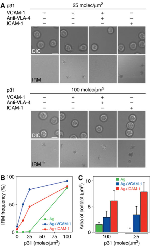
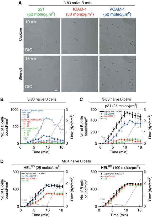
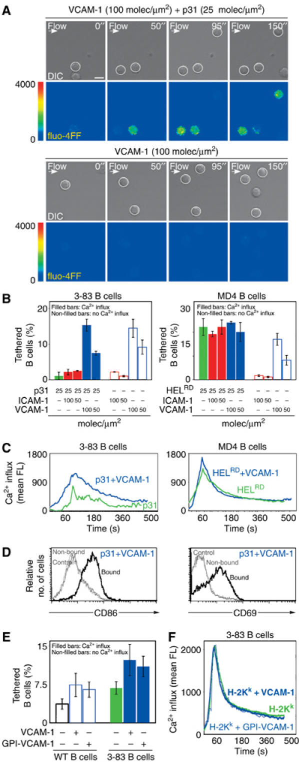
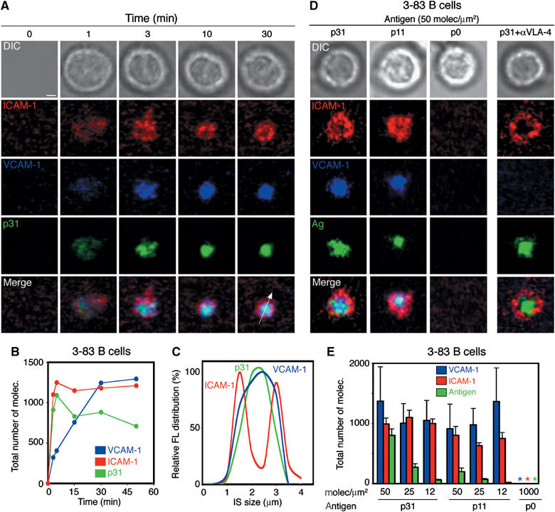
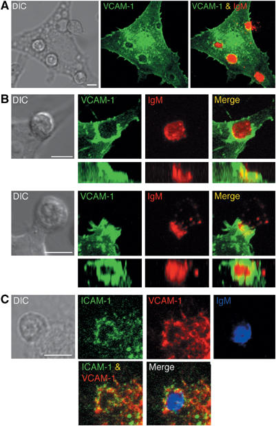
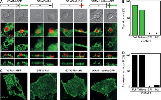
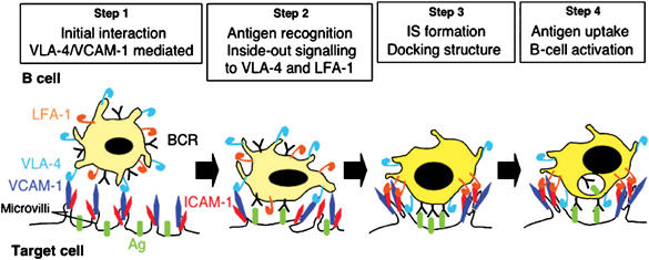
VCAM-1 is one of the main ligands of VLA-4, an integrin that is highly expressed on the surface of mature B cells. Here we find that coexpression of VCAM-1 on an antigen-bearing membrane facilitates B-cell activation. Firstly, this is achieved by mediating B-cell tethering, which in turn increases the likelihood of a B cell to be activated. Secondly, VLA-4 synergizes with the B-cell receptor (BCR), providing B cells with tight adhesion and enhanced signalling. This dual role of VCAM-1 in promoting B-cell activation is predominantly effective when the affinity of the BCR for the antigen is low. In addition, we show that the VCAM-1 ectodomain alone is sufficient to carry out this function. However, it requires the transmembrane domain to segregate properly into a docking structure characteristic of the B-cell immunological synapse (IS). These results show that the VLA-4/VCAM-1 interaction during membrane antigen recognition enhances B-cell activation and this function appears to be independent of its final peripheral localization at the IS.


Figures

Figure 1
Figure 1
VLA-4/VCAM-1 interaction increases the adhesion of B cells to target membranes. (A) Differential interference contrast (DIC) and IRM images of 3-83 naive B cells settled onto planar lipid bilayers containing p31 antigen (100 molec/μm2, top panels; 25 molec/μm2, bottom panels) in the presence or absence of ICAM-1 (50 molec/μm2), VCAM-1 (50 molec/μm2) and/or neutralizing anti-mouse α4 mAbs. Scale bar, 5 μm. (B) Naive 3-83 B cells were evaluated for their capacity to form tight contacts on membranes loaded with p31 in the presence or absence of ICAM-1 or VCAM-1 at 50 molec/μm2. After 5–10 min, images were collected at each of the specified antigen densities and the number of cells showing tight contacts was determined. Data are representative of four different experiments. (C) Quantification of the area of B-cell contact with membranes containing p31 antigen at the indicated densities in the presence or absence of ICAM-1 or VCAM-1 at 50 molec/μm2. Data represent the mean of 40 cells in each case.
Figure 2
Figure 2
VLA-4/VCAM-1 interaction facilitates the capture of naive B cells and membrane-bound antigen recognition under shear stress. (A, B) 3-83 naive B cells were perfused at decreasing shear stress for 10 min (capture step), followed by injection of buffer at increasing shear stress for 8 min (strength step). DIC images at two different time points of the same representative field of p31, ICAM-1 and VCAM-1 membranes are shown in (A). The capacity to capture B cells and the strength of binding of lipid bilayers containing VCAM-1, ICAM-1 or p31 antigen (KA=6.5 × 10⁷ M−1) at different densities (B), or of lipid bilayers containing p31 antigen in the presence or absence of VCAM-1 (50 molec/μm2) and/or ICAM-1 (50 molec/μm2) (C), were evaluated by counting B cells bound/mm2 at the indicated time points. (D) Lipid bilayers containing HELRD (KA=2 × 10⁹ M−1) at the indicated densities in the presence or absence of VCAM-1 (50 molec/μm2) and/or ICAM-1 (50 molec/μm2) were evaluated for their capacity to capture MD4 naive B cells and the strength of binding as previously described. The data in (A–D) are representative of at least three experiments. Scale bar, 20 μm.
Figure 3
Figure 3
VLA-4/VCAM-1-mediated tethering enhances B-cell activation under low shear stress. Naive B cells were perfused at low shear stress (0.1–0.05 dyn/cm2) over planar membranes containing VCAM-1 or ICAM-1 in the presence or absence of antigen. (A) Different time points of two representative experiments are shown. The upper panel in each horizontal pair shows DIC images in which tethered B cells are highlighted with a white circle. The bottom panels show their calcium response as fluo-4FF fluorescence in false colour scale. Scale bar, 5 μm. (B) The frequency of tethered B cells is represented as a percentage of the total number of perfused B cells under the different conditions analysed. The left panel shows 3-83 naive B cells on membranes loaded with the indicated densities of low-affinity p31 antigen, VCAM-1 or ICAM-1. The right panel shows the same but using MD4 B cells and HELRD as high-affinity antigen. The proportion of activated B cells was determined based on the increased levels of fluo-4FF fluorescence (filled bars: activated B cells; non-filled bars: non-activated B cells). (C) The fluo-4FF fluorescence intensity was measured every 4 s for at least 50 cells in random fields. The average intensity was plotted as a function of time for 3-83 B cells (left panel) and MD4 B cells (right panel) upon tethering on antigen-bearing membranes containing p31 and HELRD respectively, in the presence (blue line) or absence (green line) of VCAM-1. (D) Activation of the 3-83 naive B cells tethered (bound) versus the non-tethered B cells (non-bound) on membranes containing VCAM-1 and p31 as antigen. B-cell activation was evaluated after 24 h as the percentage of cells that upregulate CD86 and CD69, as described in Materials and methods. (E) As in (B), percentage of WT naive B cells or 3-83 naive B cells, which recognize H2-Kk as antigen, that tether and/or raise their calcium levels over L cells (VCAM-1+/H2-Kk), DCEK (VCAM-1/H2-Kk) or DCEK transfectants (GPI-VCAM-1+/H2-Kk). (F) As in (C), calcium response of 3-83 B cells under the conditions specified in (E).
Figure 4
Figure 4
VLA-4 can drive the recruitment of GPI-VCAM-1 to the cSMAC of the IS. Naive 3-83 B cells were settled onto planar lipid bilayers containing GPI-linked ICAM-1 (red) at 50 molec/μm2, GPI-linked VCAM-1 (blue) at 25 molec/μm2 and p31 antigen (green) at 50 molec/μm2. (A) Time-lapse fluorescent images show the accumulation of p31 (green), ICAM-1 (red) and VCAM-1 (blue) in the pattern of a mature synapse at the specified time points. The top panels show DIC images at the same time points. (B) Quantification of the total number of molecules of VCAM-1, ICAM-1 and p31 antigen during synapse formation of a representative 3-83 B cell. (C) Distribution pattern of the antigen ICAM-1 and VCAM-1 fluorescent signal across a section of the IS, shown as a white arrow in (A), at 30 min. (D) Naive 3-83 B cells were settled onto membranes containing ICAM-1 (red), VCAM-1 (blue) (50 and 25 molec/μm2, respectively) and different antigens (green) at 50 molec/μm2: p31 (KA=6.5 × 10⁷ M−1), p11 (KA∼7 × 10⁶ M−1) and p0 (null). DIC, fluorescence and merged images from a representative B cell are shown. (E) Quantification of the number of molecules of VCAM-1, ICAM-1 and antigen recruited in the B-cell synapse shown in (D). Data represent the mean of 50 cells in each case. Scale bar, 1 μm.
Figure 5
Figure 5
Full VCAM-1 localizes in the periphery of the B-cell synapse. (A) Interaction in real time of naive 3-83 B cells stained with Cy5-conjugated non-blocking anti-IgM Fabs (red) with L cells expressing VCAM-1-GFP (green) was analysed by confocal microscopy. Separate and merged three-dimensional projections of the confocal fluorescence image stacks taken after 20 min incubation are shown. DIC image at the same time point is shown in the left panel. (B) Side and top views of the three-dimensional reconstruction of the docking structure formed by VCAM-1-GFP in the B-cell synapse. Two different examples of DIC, separate and merged fluorescent images of VCAM-1-GFP and IgM are shown. (C) 3-83 B cells interacting with ICAM-1-GFP target cells were fixed, permeabilized and stained for endogenous VCAM-1 and IgM with anti-VCAM-1 (red) and anti-IgM antibodies (blue). DIC, separate and merged three-dimensional projection of the confocal fluorescence image stacks of VCAM-1, ICAM-1 and IgM are shown. Scale bar, 5 μm.
Figure 6
Figure 6
The transmembrane domain of VCAM-1 determines its distribution in the IS. (A) Interaction of 3-83 naive B cells with DCEK cells transfected with the different VCAM-1 constructs. 3-83 B cells interacting with DCEK cells transfected with GPI-VCAM-1 or EC VCAM-1/H2 were fixed, permeabilized and stained with antibodies against VCAM-1 and IgM, as indicated in Materials and methods. 3-83 B cells, previously stained with Cy5-conjugated non-blocking anti-IgM Fabs, were in contact with VCAM-1-GFP and VCAM-1 tailless-GFP DCEK transfectants for 25 min at 37°C. Then, confocal life-imaging was acquired. Two different examples are shown for each VCAM-1 construct. The middle panels show top views of fluorescent images representing a projection of several horizontal confocal sections. The bottom panels show a sagittal section of the top view in each case. DIC images are shown in the top panel. Schematic illustrations of the different VCAM-1 constructs are shown on top of the corresponding panels. Scale bar, 5 μm. (B) The four VCAM-1 constructs were evaluated for their capacity to be excluded and form a docking structure in the B-cell synapse. Data obtained from at least 30 B-cell synapses were analysed in each case. (C) COS transfectants of the different VCAM-1 constructs were fixed and stained with anti-VCAM-1 mAb (green). Each panel represents a projection of the confocal fluorescence image stack. Scale bar, 5 μm. (D) The four VCAM-1 constructs were evaluated for their capacity to be expressed on microvilli at the surface of DCEK cells. Data obtained from at least 30 DCEK transfected cells were analysed in each case.
Figure 7
Figure 7
Model of B-cell membrane antigen recognition.

Similar articles

Cited by

References

    1. Alon R, Kassner PD, Carr MW, Finger EB, Hemler ME, Springer TA (1995) The integrin VLA-4 supports tethering and rolling in flow on VCAM-1. J Cell Biol 128: 1243–1253 - PMC - PubMed
    1. Atherton A, Born GV (1972) Quantitative investigations of the adhesiveness of circulating polymorphonuclear leucocytes to blood vessel walls. J Physiol 222: 447–474 - PMC - PubMed
    1. Barreiro O, Yanez-Mo M, Sala-Valdes M, Gutierrez-Lopez MD, Ovalle S, Higginbottom A, Monk PN, Cabanas C, Sanchez-Madrid F (2005) Endothelial tetraspanin microdomains regulate leukocyte firm adhesion during extravasation. Blood 105: 2852–2861 - PubMed
    1. Barreiro O, Yanez-Mo M, Serrador JM, Montoya MC, Vicente-Manzanares M, Tejedor R, Furthmayr H, Sanchez-Madrid F (2002) Dynamic interaction of VCAM-1 and ICAM-1 with moesin and ezrin in a novel endothelial docking structure for adherent leukocytes. J Cell Biol 157: 1233–1245 - PMC - PubMed
    1. Batista FD, Neuberger MS (1998) Affinity dependence of the B cell response to antigen: a threshold, a ceiling, and the importance of off-rate. Immunity 8: 751–759 - PubMed

Publication types