. 2005;7(6):R1058-79.
doi: 10.1186/bcr1343. Epub 2005 Nov 8.

HER-2 overexpression differentially alters transforming growth factor-beta responses in luminal versus mesenchymal human breast cancer cells

Affiliations

HER-2 overexpression differentially alters transforming growth factor-beta responses in luminal versus mesenchymal human breast cancer cells

Cindy A Wilson et al. Breast Cancer Res. 2005.

Abstract

Introduction: Amplification of the HER-2 receptor tyrosine kinase has been implicated in the pathogenesis and aggressive behavior of approximately 25% of invasive human breast cancers. Clinical and experimental evidence suggest that aberrant HER-2 signaling contributes to tumor initiation and disease progression. Transforming growth factor beta (TGF-beta) is the dominant factor opposing growth stimulatory factors and early oncogene activation in many tissues, including the mammary gland. Thus, to better understand the mechanisms by which HER-2 overexpression promotes the early stages of breast cancer, we directly assayed the cellular and molecular effects of TGF-beta1 on breast cancer cells in the presence or absence of overexpressed HER-2.

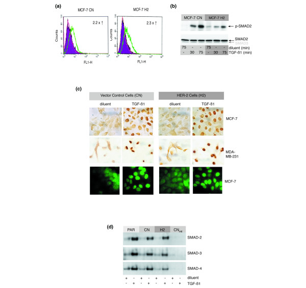
Methods: Cell proliferation assays were used to determine the effect of TGF-beta on the growth of breast cancer cells with normal or high level expression of HER-2. Affymetrix microarrays combined with Northern and western blot analysis were used to monitor the transcriptional responses to exogenous TGF-beta1 in luminal and mesenchymal-like breast cancer cells. The activity of the core TGF-beta signaling pathway was assessed using TGF-beta1 binding assays, phospho-specific Smad antibodies, immunofluorescent staining of Smad and Smad DNA binding assays.

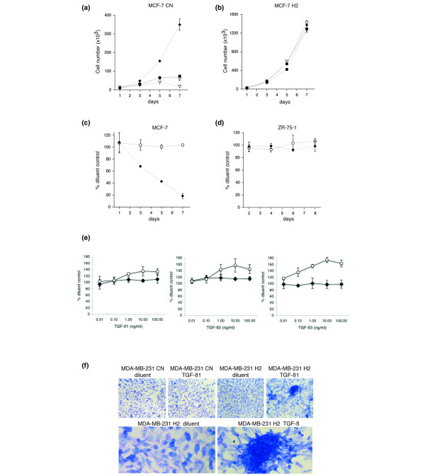
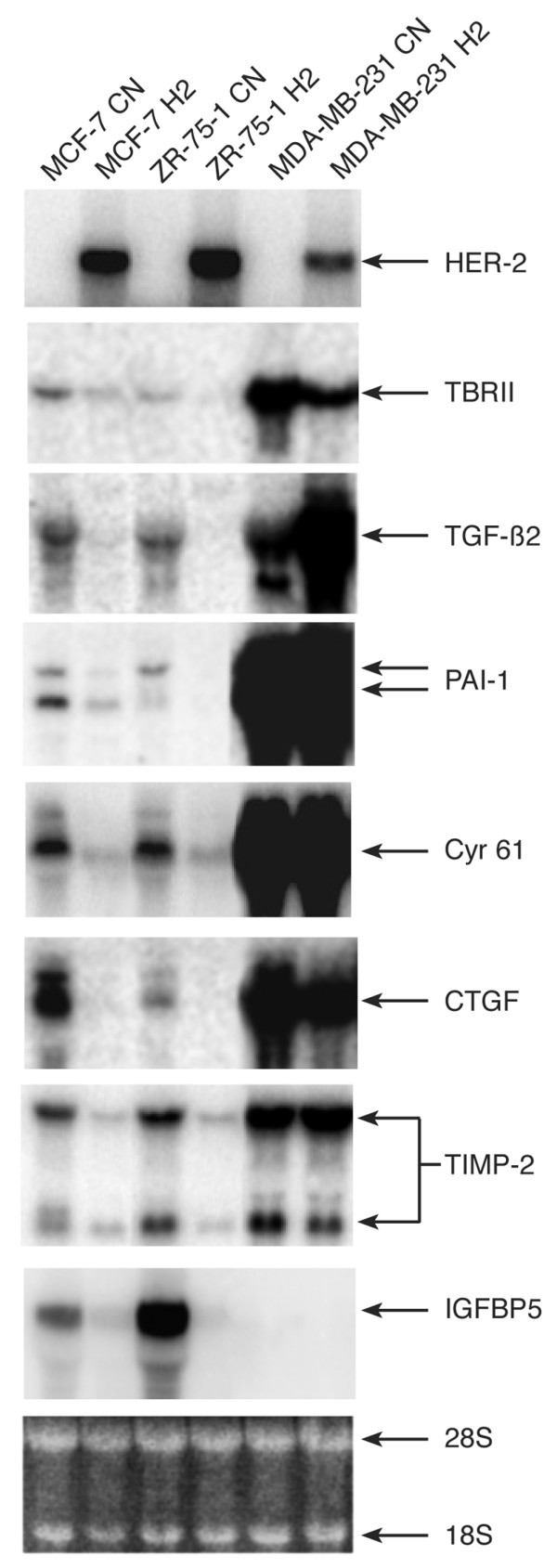
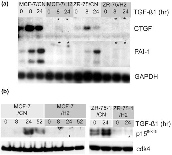
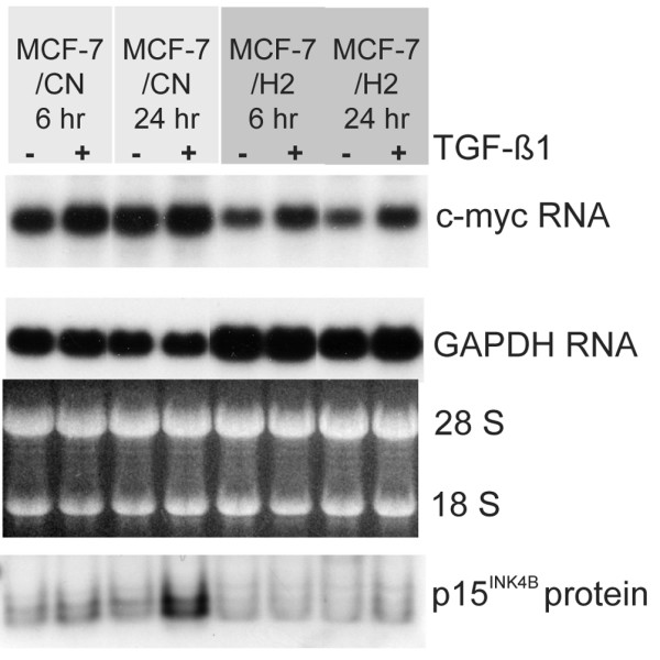
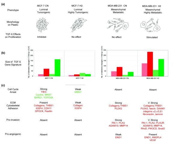
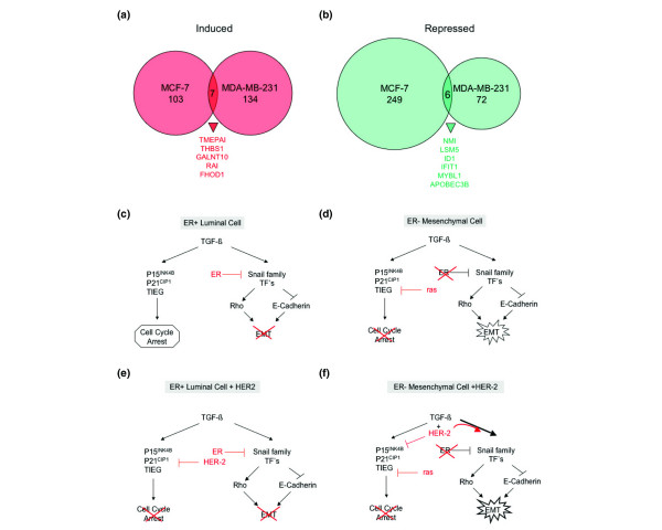
Results: We demonstrate that cells engineered to over-express HER-2 are resistant to the anti-proliferative effect of TGF-beta1. HER-2 overexpression profoundly diminishes the transcriptional responses induced by TGF-beta in the luminal MCF-7 breast cancer cell line and prevents target gene induction by a novel mechanism that does not involve the abrogation of Smad nuclear accumulation, DNA binding or changes in c-myc repression. Conversely, HER-2 overexpression in the context of the mesenchymal MDA-MB-231 breast cell line potentiated the TGF-beta induced pro-invasive and pro-metastatic gene signature.

Conclusion: HER-2 overexpression promotes the growth and malignancy of mammary epithelial cells, in part, by conferring resistance to the growth inhibitory effects of TGF-beta. In contrast, HER-2 and TGF-beta signaling pathways can cooperate to promote especially aggressive disease behavior in the context of a highly invasive breast tumor model.


Figures

Figure 1
Figure 1
HER-2 overexpression modulates transforming growth factor (TGF)-β responses in a cell context dependent manner. (a-c) HER-2 overexpression in MCF-7 breast cancer cells blocks TGF-β mediated growth arrest. CN and H2 cells were treated with diluent control (diamonds) or TGF-β1 (0.2 ng/ml (black circles); 0.4 ng/ml (white circles); or 0.8 ng/ml (triangles)) and counted on the indicated days after treatment. Each point is the average of triplicate wells (± standard deviation for error bars). (a) The growth of MCF-7 CN cells is severely inhibited by TGF-β1. (b) MCF-7 H2 cells are resistant to growth inhibition by TGF-β1. (c) MCF-7 CN (diamonds) versus MCF-7 H2 (white circles) cells treated with 0.2 ng/ml TGF-β1. (d) ZR-75-1 cells are resistant to growth inhibition by TGF-β1 without HER-2 overexpression (ZR-75-1/CN (diamonds) versus ZR-75-1/H2 (white circles)) treated with 0.2 ng/ml TGF-β1. (e) TGF-β1 stimulates the growth of MDA-MB-231 H2 cells. MDA MB-231 CN (diamonds) and H2 cells (white cirlcles) were grown for 6 days in the presence of TGF-β1, β2 or β3 (0.01 to 100 ng/ml) or a diluent control. Cells were pulsed with [3H] thymidine for the final 24 h of assay and thymidine incorporation was measured. The average counts of triplicate wells for each data point are represented as % of diluent control. (f) TGF-β induces a 'piling' phenotype in MDA-MB-231 that is dependent on HER-2 overexpression. MDA-MB-231 CN and H2 cells were grown for 5 days in the presence of 10 ng/ml TGF-β1 or diluent control. Cells were stained with crystal violet dye and photographed with a 20× (top four panels) or a 60× (bottom two panels) objective.
Figure 2
Figure 2
Expression of TGF-β pathway genes in control (CN) and HER-2 overexpressing (H2) breast cancer cells. Northern analysis of HER-2 mRNA and transforming growth factor (TGF)-β markers in MCF-7, ZR-75-1 and MDA-MB-231 cells with (H2) and without (CN) HER-2 overexpression. Message levels were assayed from 10 ug of total RNA. Asterisks indicate consistent and significant down-regulation (> 2-fold as measured by phosphorimager quantification). The image of the 28S and 18S ribosomal RNA bands from the ethidium bromide stained gel is shown as a loading control.
Figure 3
Figure 3
HER-2 overexpression inhibits transforming growth factor (TGF)-β1 target gene induction in the luminal MCF-7 and ZR-75-1 breast cancer cells. (a) Northern blot analysis of CTGF and PAI-1 expression. The membrane contained total RNA from the indicated CN and H2 cells treated with either diluent control (0 h) or 2 ng/ml TGF-β1 (8 or 24 h). (b) HER-2 overexpression prevents p15INK4B induction in MCF-7 and ZR-75-1 cells by TGF-β1. Western blot analysis of total cellular extracts prepared after treatment with diluent control (0 h) or 2 ng/ml TGF-β1 (8, 24 or 52 h). Bottom panels are the same membranes re-probed with an antibody to cdk4.
Figure 4
Figure 4
Context specific effects of HER-2 overexpression on the biological responses and transcription program induced by transforming growth factor (TGF)-β in breast cancer cells. (a) Phenotypes of the luminal MCF-7 and mesenchymal MDA-MB-231 cells with and without engineered HER-2 overexpression. The epithelial growth pattern of MCF-7 cells is characteristically altered by HER-2 overexpression, which promotes an elongated morphology and increased proliferation rates in vitro as well as faster growing tumor xenografts in vivo [12,13]. The MDA-MB-231 cells are heterogeneous with the majority of cells, having a spindle shaped morphology. The effect of HER-2 overexpression on the morphological appearance was not dramatic except that significantly more (approximately three times) of the large, flattened round cells were observed. The MDA-MB-231 H2 cells have also been shown to be more metastatic in vivo than the MDA-MB-231 CN or parental lines (data not shown). (b) Summary of the TGF-β transcriptional program obtained by Affymetrix microarray profiling of cells treated with recombinant TGF-β1. The number of elements significantly affected (p < 0.01 and fold change greater than ± 1.7 using Rosetta Resolver) are graphed. Light and dark red indicate genes upregulated after 6 h and 24 h, respectively, and light and dark green represent genes downregulated after 6 and 24 h, respectively. (c) Highlighted genes from the dominant functional gene signatures as determined by the gene ontology information found in Source [105] and GeneCards [106]. Red and green indicate TGF-β stimulated and repressed genes, respectively. ECM, extracellular matrix.
Figure 5
Figure 5
HER-2 overexpression does not alter transforming growth factor (TGF)-β1 induced Smad2 activation or nuclear accumulation or the formation of Smad-Smad-binding element (SBE) complexes. (a) TGF-β1 binds equally well to the surface of MCF-7 CN and MCF-7 H2 cells. The binding of fluorescein isothiocyanate (FITC)-labeled recombinant TGF-β1 was monitored by flow cytometry. The red curves show the FL1 values for the untreated control cells, the purple (filled) curves indicate the intensity of cells incubated with an irrelevant FITC-labeled protein and the green curves show the intensity of cells treated with FITC-labeled recombinant TGF-β1. The median shift in FL1 values (fold increase) between the irrelevant control protein and TGF-β1 is indicated. (b) TGF-β1 induced phosphorylation of Smad2 is detected in both MCF-7 CN and MCF-7 H2 cells. A phospho-specific Smad2 antibody was used on immunoblots of whole cell lysates from diluent or TGF-β1 treated cells. The blot was re-probed with an antibody against total Smad2, which cross-reacts weakly with Smad3. (c) Endogenous Smad2 is concentrated in the nucleus after exposure to TGF-β1 in MCF-7 or MDA-MB-231 cells with or without HER-2 overexpression. Vector control (CN) cells (left panels) or HER-2 overexpressing (H2) cells (right panels) were treated for 1 h with either diluent control or 2 ng/ml TGF-β1, fixed and stained with the anti-Smad2 antibody. The endogenous Smad2 protein was visualized with either a peroxidase/3,3'-Diaminobenzidine (DAB) reaction (top two rows) or an Alexa-488 conjugated secondary antibody (bottom row). (d) TGF-β1 stimulated Smad DNA binding is not affected by HER-2 overexpression in MCF-7 cells. Nuclear protein extracts from MCF-7 parental (PAR), MCF-7 CN and MCF-7 H2 cells treated with either diluent or TGF-β1 were reacted with biotin labeled oligonucleotides containing SBEs. Avidin-coupled sepharose beads were used to collect the DNA-protein complexes. The Smad composition of the complexes was analyzed by western blotting with anti-Smad antibodies as indicated. The last two lanes (CNmt) contained the same MCF-7 CN nuclear extract as in lanes 3–4, with a mutant oligonucleotide in which one of the SBE sites was mutated (i.e. PE2Sm1 [41]).
Figure 6
Figure 6
Downregulation of c-myc RNA levels by transforming growth factor (TGF)-β is not observed and is not required for p15INK4B induction in MCF-7 cells. RNA and total protein were extracted from the same flasks of cells treated for either 6 or 24 h with diluent control or 2 ng/ml TGF-β1. The protein samples were probed with an anti-p15INK4B antibody as in Fig. 4b. The Northern blot re-probed with a glyceraldehyde-3-phosphate dehydrogenase (GAPDH) sequence and the image of the 28S and 18S ribosomal RNAs serve as controls.
Figure 7
Figure 7
Divergent transforming growth factor (TGF)-β gene activation and biological responses in MCF-7 and MDA-MB-231 cell lines. (a,b) The TGF-β induced transcriptional program is almost entirely non-overlapping in the luminal MCF-7 and the mesenchymal MDA-MB-231 breast cancer cell lines. The number of genes (a) induced and (b) repressed greater than 1.5-fold with a p-value < 0.01 after a 24 h exposure to TGF-β are shown. All of the individual genes commonly affected in both control cell lines (MCF-7 CN and MDA-MB-231 CN) are listed below the diagram. (c,d) Model for the differential outcomes of TGF-β stimulation in estrogen receptor (ER) positive luminal breast cells compared to ER-negative mesenchymal cells. (e,f) Model for context specific effects of HER-2 overexpression on TGF-β signaling in ER-positive luminal cells versus ER-negative mesenchymal cells. EMT, epithelial to mesenchymal transition.

Similar articles

Cited by

References

    1. King CR, Kraus MH, Aaronson SA. Amplification of a novel v-erbB-related gene in a human mammary carcinoma. Science. 1985;229:974–976. - PubMed
    1. Coussens L, Yang-Feng TL, Liao YC, Chen E, Gray A, McGrath J, Seeburg PH, Libermann TA, Schlessinger J, Francke U, et al. Tyrosine kinase receptor with extensive homology to EGF receptor shares chromosomal location with neu oncogene. Science. 1985;230:1132–1139. - PubMed
    1. Tzahar E, Waterman H, Chen X, Levkowitz G, Karunagaran D, Lavi S, Ratzkin BJ, Yarden Y. A hierarchical network of interreceptor interactions determines signal transduction by Neu differentiation factor/neuregulin and epidermal growth factor. Mol Cell Biol. 1996;16:5276–5287. - PMC - PubMed
    1. Graus-Porta D, Beerli RR, Daly JM, Hynes NE. ErbB-2, the preferred heterodimerization partner of all ErbB receptors, is a mediator of lateral signaling. EMBO J. 1997;16:1647–1655. doi: 10.1093/emboj/16.7.1647. - DOI - PMC - PubMed
    1. Slamon DJ, Clark GM, Wong SG, Levin WJ, Ullrich A, McGuire WL. Human breast cancer: correlation of relapse and survival with amplification of the HER-2/neu oncogene. Science. 1987;235:177–182. - PubMed

Publication types

Substances