. 2006 Feb 3:4:6.
doi: 10.1186/1477-7827-4-6.

Two unrelated putative membrane-bound progestin receptors, progesterone membrane receptor component 1 (PGMRC1) and membrane progestin receptor (mPR) beta, are expressed in the rainbow trout oocyte and exhibit similar ovarian expression patterns

Affiliations

Two unrelated putative membrane-bound progestin receptors, progesterone membrane receptor component 1 (PGMRC1) and membrane progestin receptor (mPR) beta, are expressed in the rainbow trout oocyte and exhibit similar ovarian expression patterns

Brigitte Mourot et al. Reprod Biol Endocrinol. .

Abstract

Background: In lower vertebrates, steroid-induced oocyte maturation is considered to involve membrane-bound progestin receptors. Two totally distinct classes of putative membrane-bound progestin receptors have been reported in vertebrates. A first class of receptors, now termed progesterone membrane receptor component (PGMRC; subtypes 1 and 2) has been studied since 1996 but never studied in a fish species nor in the oocyte of any animal species. A second class of receptors, termed membrane progestin receptors (mPR; subtypes alpha, beta and gamma), was recently described in vertebrates and implicated in the progestin-initiated induction of oocyte maturation in fish.

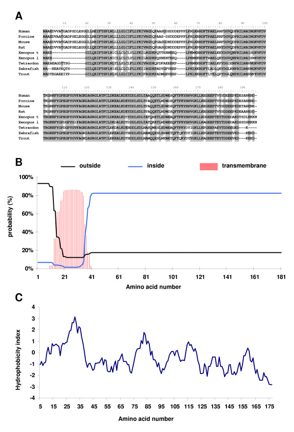
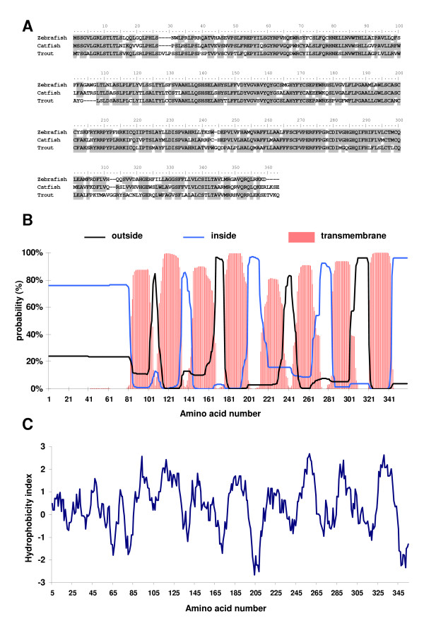
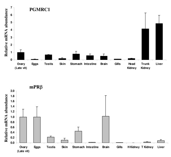
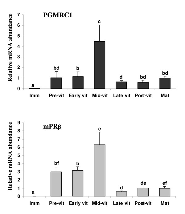
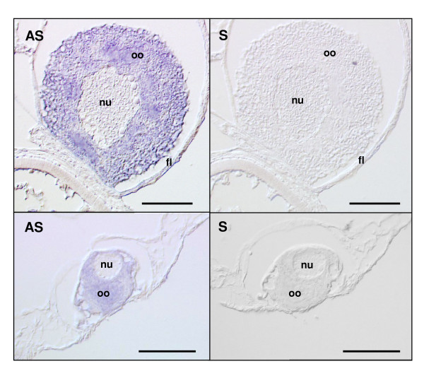
Methods: In the present study, we report the characterization of the full coding sequence of rainbow trout PGMRC1 and mPR beta cDNAs, their tissue distribution, their ovarian expression profiles during oogenesis, their hormonal regulation in the full grown ovary and the in situ localization of PGMRC1 mRNA in the ovary.

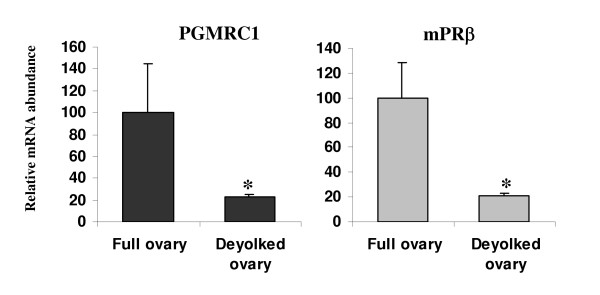
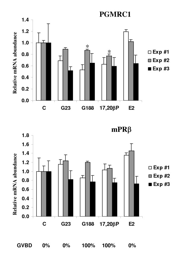
Results: Our results clearly show, for the first time in any animal species, that rainbow trout PGMRC1 mRNA is present in the oocyte and has a strong expression in ovarian tissue. In addition, we show that both mPR beta and PGMRC1, two members of distinct membrane-bound progestin receptor classes, exhibit highly similar ovarian expression profiles during the reproductive cycle with maximum levels during vitellogenesis and a down-expression during late vitellogenesis. In addition, the mRNA abundance of both genes is not increased after in vitro hormonal stimulation of full grown follicles by maturation inducing hormones.

Conclusion: Together, our findings suggest that PGMRC1 is a new possible participant in the progestin-induced oocyte maturation in fish. However, its participation in the process of oocyte maturation, which remains to be confirmed, would occur at post-transcriptional levels.


Figures

Figure 1
Figure 1
(A) Alignment of the deduced amino acid sequence of rainbow trout PGMRC1 with other vertebrates PGMRC1 and PGMRC2 proteins. Most conserved residues are shaded. Corresponding GenBank accession numbers are shown in Table 1. (B) Outside, inside and transmembrane probability of rainbow trout PGMRC1 deduced protein. (C) Hydrophobicity analysis of rainbow trout PGMRC1 deduced protein sequence according to Kyte and Doolittle [36].
Figure 2
Figure 2
Alignment of rainbow trout (ABA39295), zebrafish (NP_899187) and catfish (AAS45555) mPRβ deduced amino acid sequences. Most conserved residues are shaded. (B) Outside, inside and transmembrane probability of rainbow trout mPRβ deduced protein. (C) Hydrophobicity analysis of rainbow trout mPRβ deduced protein sequence according to Kyte and Doolittle [36].
Figure 3
Figure 3
Tissue distribution of PGMRC1 (A) and mPRβ (B) mRNAs in rainbow trout. Tissues were collected from 3 different females during reproductive period. Ovaries were sampled at late vitellogenic stage. Testes were sampled at stage II of spermatogenesis [31]. The mRNA abundance (mean ± SEM, N = 3 tissue samples originating from different individuals) of each gene was determined by real-time PCR and normalized to the abundance of 18S. For each gene, mRNA abundance was arbitrarily set to 1 for the "ovary" group.
Figure 4
Figure 4
Ovarian expression profiles of PGMRC1 and mPRβ mRNAs during rainbow trout oogenesis (mean ± SEM). Ovaries were sampled from separate females during immature stage (Imm, N = 5 fish, 1000°C. days post-fertilization), pre-vitellogenesis (Pre-vit, N = 6 fish), early vitellogenesis (early vit, N = 6 fish), mid-vitellogenesis (Mid-vit, N = 6 fish), late vitellogenesis (late vit, N = 6 fish), post-vitellogenesis (post-vit, N = 6 fish) and oocyte maturation (Mat, N = 4 fish). The mRNA abundance (mean ± SEM) of each gene was determined by real-time PCR and normalized to the abundance of 18S. For each gene, mRNA abundance was arbitrarily set to 1 for oocyte maturation group. Different letters indicate significant differences between groups (p < 0.05).
Figure 5
Figure 5
Comparative mRNA abundance of PGMRC1 and mPRβ mRNAs in the rainbow trout intact (N = 3 groups of 25 follicles) or deyolked (N = 3 groups of 25 follicles) post-vitellogenic ovary. The mRNA abundance of each gene (mean ± SEM) was determined by real-time PCR and normalized to the abundance of 18S. For each gene, mRNA abundance was arbitrarily set to 1 in the intact ovary. Asterisks indicate significant differences (p < 0.05).
Figure 6
Figure 6
Hormonal regulation of mPRβ and PGMRC1 mRNA abundance in full-grown intact rainbow trout ovarian follicles. The experiment was repeated 3 times and for each experiment, all follicles originated from the same ovary. For each hormonal treatment (C = Control; 17,20βP = 17,20β-dihydroxy-4pregnen-3one, 40 ng/ml; G23, G188 = partially purified gonadotropins, 23 or 188 ng/ml; E2 = estradiol, 1 μg/ml) the 20 h incubation was performed in triplicates. The mRNA abundance (mean ± SEM, N = 3 groups of 25 follicles) of each gene was determined by real-time PCR and normalized to the abundance of 18 S. For each experiment, the mRNA abundance of each gene was arbitrarily set to 1 for the control group. *: significantly different from the control group of the experiment at p < 0.05. For each hormonal treatment, occurrence of germinal vesicle breakdown (GVBD) observed after 60 h is indicated below the graphs.
Figure 7
Figure 7
In situ hybridization of mid-vitellogenic ovarian tissue in the presence or PGMRC1 antisense (AS) and sense (S) riboprobes. Positive (AS) and background (S) staining of a previtellogenic oocyte are shown. Labels:n = nucleus, oo = ooplasm, fl = follicle somatic layers. Bars represent 50 μm.

References

    1. O'Malley BW. A life-long search for the molecular pathways of steroid hormone action. Mol Endocrinol. 2005;19:1402–1411. doi: 10.1210/me.2004-0480. - DOI - PubMed
    1. Falkenstein E, Tillmann HC, Christ M, Feuring M, Wehling M. Multiple actions of steroid hormones--a focus on rapid, nongenomic effects. Pharmacol Rev. 2000;52:513–556. - PubMed
    1. Pietras RJ, Szego CM. Specific binding sites for oestrogen at the outer surfaces of isolated endometrial cells. Nature. 1977;265:69–72. doi: 10.1038/265069a0. - DOI - PubMed
    1. Smith LD, Ecker RE. Role of the oocyte nucleus in physiological maturation in Rana pipiens. Dev Biol. 1969;19:281–309. doi: 10.1016/0012-1606(69)90065-7. - DOI - PubMed
    1. Zhu Y, Bond J, Thomas P. Identification, classification, and partial characterization of genes in humans and other vertebrates homologous to a fish membrane progestin receptor. Proc Natl Acad Sci U S A. 2003;100:2237–2242. doi: 10.1073/pnas.0436133100. - DOI - PMC - PubMed

LinkOut - more resources