

Regulation of p90RSK phosphorylation by SARS-CoV infection in Vero E6 cells
- PMID: 16458888
- PMCID: PMC7094696
- DOI: 10.1016/j.febslet.2006.01.066
Regulation of p90RSK phosphorylation by SARS-CoV infection in Vero E6 cells
Abstract
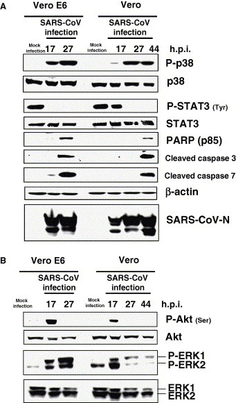
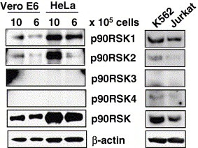
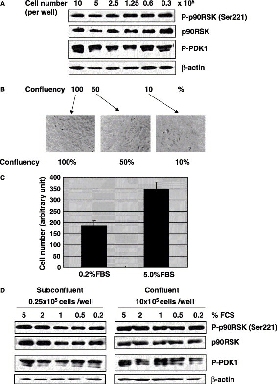
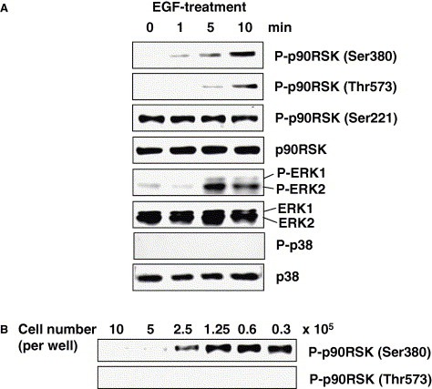
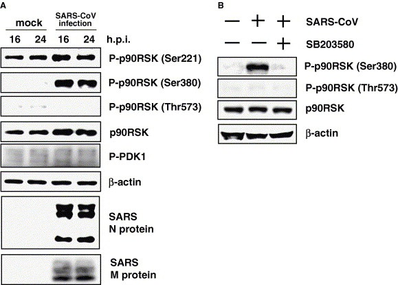
The 90 kDa ribosomal S6 kinases (p90RSKs) are a family of broadly expressed serine/threonine kinases with two kinase domains activated by extracellular signal-regulated protein kinase in response to many growth factors. Our recent study demonstrated that severe acute respiratory syndrome (SARS)-coronavirus (CoV) infection of monkey kidney Vero E6 cells induces phosphorylation and dephosphorylation of signaling pathways, resulting in apoptosis. In the present study, we investigated the phosphorylation status of p90RSK, which is a well-known substrate of these signaling pathways, in SARS-CoV-infected cells. Vero E6 mainly expressed p90RSK1 and showed weak expression of p90RSK2. In the absence of viral infection, Ser221 in the N-terminal kinase domain was phosphorylated constitutively, whereas both Thr573 in the C-terminal kinase domain and Ser380 between the two kinase domains were not phosphorylated in confluent cells. Ser380, which has been reported to be involved in autophosphorylation by activation of the C-terminal kinase domain, was phosphorylated in confluent SARS-CoV-infected cells, and this phosphorylation was inhibited by , which is an inhibitor of p38 mitogen-activated protein kinases (MAPK). Phosphorylation of Thr573 was not upregulated in SARS-CoV-infected cells. Thus, in virus-infected cells, phosphorylation of Thr573 was not necessary to induce phosphorylation of Ser380. On the other hand, Both Thr573 and Ser380 were phosphorylated by treatment with epidermal growth factor (EGF) in the absence of p38 MAPK activation. Ser220 was constitutively phosphorylated despite infection. These results indicated that phosphorylation status of p90RSK by SARS-CoV infection is different from that by stimulation of EGF. This is the first detailed report regarding regulation of p90RSK phosphorylation by virus infection.
Figures
Similar articles
-
Tyrosine dephosphorylation of STAT3 in SARS coronavirus-infected Vero E6 cells.FEBS Lett. 2004 Nov 5;577(1-2):187-92. doi: 10.1016/j.febslet.2004.10.005. FEBS Lett. 2004. PMID: 15527783 Free PMC article.
-
Inhibition of cell proliferation by SARS-CoV infection in Vero E6 cells.FEMS Immunol Med Microbiol. 2006 Mar;46(2):236-43. doi: 10.1111/j.1574-695X.2005.00028.x. FEMS Immunol Med Microbiol. 2006. PMID: 16487305 Free PMC article.
-
JNK and PI3k/Akt signaling pathways are required for establishing persistent SARS-CoV infection in Vero E6 cells.Biochim Biophys Acta. 2005 Jun 30;1741(1-2):4-10. doi: 10.1016/j.bbadis.2005.04.004. Biochim Biophys Acta. 2005. PMID: 15916886 Free PMC article.
-
Signal transduction in SARS-CoV-infected cells.Ann N Y Acad Sci. 2007 Apr;1102(1):86-95. doi: 10.1196/annals.1408.006. Ann N Y Acad Sci. 2007. PMID: 17470913 Free PMC article. Review.
-
Role of p90RSK in Kidney and Other Diseases.Int J Mol Sci. 2019 Feb 23;20(4):972. doi: 10.3390/ijms20040972. Int J Mol Sci. 2019. PMID: 30813401 Free PMC article. Review.
Cited by
-
Lysophosphatidic Acid Triggers Apoptosis in HeLa Cells through the Upregulation of Tumor Necrosis Factor Receptor Superfamily Member 21.Mediators Inflamm. 2017;2017:2754756. doi: 10.1155/2017/2754756. Epub 2017 Feb 19. Mediators Inflamm. 2017. PMID: 28348459 Free PMC article.
-
A persistently infecting coronavirus in hibernating Myotis lucifugus, the North American little brown bat.J Gen Virol. 2017 Sep;98(9):2297-2309. doi: 10.1099/jgv.0.000898. Epub 2017 Aug 25. J Gen Virol. 2017. PMID: 28840816 Free PMC article.
-
Enhancement of cytotoxicity against Vero E6 cells persistently infected with SARS-CoV by Mycoplasma fermentans.Arch Virol. 2007;152(5):1019-25. doi: 10.1007/s00705-006-0924-7. Epub 2007 Feb 7. Arch Virol. 2007. PMID: 17277901 Free PMC article.
-
Molecular pathogenesis of severe acute respiratory syndrome.Microbes Infect. 2007 Jan;9(1):119-26. doi: 10.1016/j.micinf.2006.06.012. Epub 2006 Sep 28. Microbes Infect. 2007. PMID: 17142081 Free PMC article. Review.
-
Combination Kinase Inhibitor Treatment Suppresses Rift Valley Fever Virus Replication.Viruses. 2018 Apr 13;10(4):191. doi: 10.3390/v10040191. Viruses. 2018. PMID: 29652799 Free PMC article.
References
-
- Pearson G., Robinson F., Beers Gibson T., Xu B.E., Karandikar M., Berman K., Cobb M.H., Mitogen-activated protein (MAP) kinase pathways: regulation and physiological functions. Endocr. Rev., 22, (2001), 153– 183. - PubMed
-
- Frodin M., Gammeltoft S., Role and regulation of 90 kDa ribosomal S6 kinase (RSK) in signal transduction. Mol. Cell. Endocrinol., 151, (1999), 65– 77. - PubMed
-
- Moller D.E., Xia C.H., Tang W., Zhu A.X., Jakubowski M., Human rsk isoforms: cloning and characterization of tissue-specific expression. Am. J. Physiol., 266, (1994), C351– C359. - PubMed
-
- Yntema H.G., van den Helm B., Kissing J., van Duijnhoven G., Poppelaars F., Chelly J., Moraine C., Fryns J.P., Hamel B.C., Heilbronner H., Pander H.J., Brunner H.G., Ropers H.H., Cremers F.P., van Bokhoven H., A novel ribosomal S6-kinase (RSK4; RPS6KA6) is commonly deleted in patients with complex X-linked mental retardation. Genomics, 62, (1999), 332– 343. - PubMed
Publication types
MeSH terms
Substances
LinkOut - more resources
Full Text Sources
Other Literature Sources
Molecular Biology Databases
Miscellaneous
