

Young-elderly differences in bone density, geometry and strength indices depend on proximal femur sub-region: a cross sectional study in Caucasian-American women
- PMID: 16459156
- PMCID: PMC1482801
- DOI: 10.1016/j.bone.2005.11.020
Young-elderly differences in bone density, geometry and strength indices depend on proximal femur sub-region: a cross sectional study in Caucasian-American women
Abstract
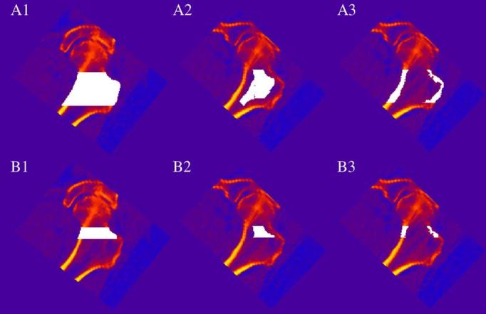
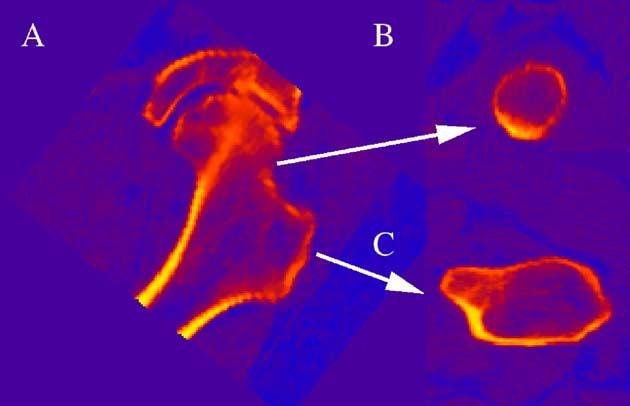
Fragility fractures at the trochanter (TR) and the femoral neck (FN) have distinct etiologies, but the underlying age-related structural changes at these proximal femoral sub-regions are poorly understood. 28 young (41+/-3 years) and 124 elderly (74+/-3 years) healthy Caucasian women underwent volumetric quantitative computed tomography at the hip. Integral (i), cortical (c) and trabecular (t) bone mineral density and content (BMD, BMC) were measured. Geometric parameters included cross sectional area (CSA), and volumes of the integral, cortical and trabecular regions (VOL). Structural measures included indices of compressive (Compstr) and bending (BSI) strength. After adjusting for height and weight, an F-test was used to compare the TR and the FN mean values between young and elderly and to test for interaction to compare logarithmic difference of young and elderly (log(Young)-log(Elderly), Y/Ed) between the FN and the TR in an ANOCOVA model. All BMC, iBMD and tBMD values were significantly lower in elderly than in young women, with the largest Y/Ed in the FN tBMC and tBMD (P<0.0011 and P<0.0001). cBMD in young and elderly groups was not significantly different at the TR while at the FN it was greater (P=0.0075) in elderly than young women, showing significant Y/Ed (P=0.0003) dependence on skeletal site. Elderly women had significantly larger iVOL and CSA values (0.0001<P<0.0051), except for the FN iVOL. cVOL values were smaller in elderly than young women (P<0.0001). Y/Ed in bone geometry differed by sub-region only for cVOL measures (P=0.0267). Despite larger CSA and iVOL measures in elderly, the younger women had greater Compstr (P<0.0001) and BSI (P=0.0051). Thus, although both the TR and the FN appear to increase in size with age, this enlargement is insufficient to protect against loss of bone strength.
Figures
Similar articles
-
Structural and biomechanical basis of sexual dimorphism in femoral neck fragility has its origins in growth and aging.J Bone Miner Res. 2003 Oct;18(10):1766-74. doi: 10.1359/jbmr.2003.18.10.1766. J Bone Miner Res. 2003. PMID: 14584886
-
Age-related loss of proximal femoral strength in elderly men and women: the Age Gene/Environment Susceptibility Study--Reykjavik.Bone. 2012 Mar;50(3):743-8. doi: 10.1016/j.bone.2011.12.001. Epub 2011 Dec 10. Bone. 2012. PMID: 22178403 Free PMC article.
-
Effects of gender, anthropometric variables, and aging on the evolution of hip strength in men and women aged over 65.Bone. 2003 May;32(5):561-70. doi: 10.1016/s8756-3282(03)00055-3. Bone. 2003. PMID: 12753873
-
Quantitative computed tomography reveals the effects of race and sex on bone size and trabecular and cortical bone density.J Clin Densitom. 2009 Jul-Sep;12(3):330-6. doi: 10.1016/j.jocd.2009.04.001. Epub 2009 Jul 4. J Clin Densitom. 2009. PMID: 19577939
-
Varying contributions of growth and ageing to racial and sex differences in femoral neck structure and strength in old age.Bone. 2005 Jun;36(6):978-86. doi: 10.1016/j.bone.2004.11.015. Bone. 2005. PMID: 15869919
Cited by
-
The rate of hip osteoarthritis in patients with proximal femoral fractures versus hip contusion.Osteoporos Int. 2012 Mar;23(3):901-5. doi: 10.1007/s00198-011-1628-8. Epub 2011 Apr 6. Osteoporos Int. 2012. PMID: 21625883 Free PMC article.
-
Effects of age and sex on the strength and cortical thickness of the femoral neck.Bone. 2011 Apr 1;48(4):741-7. doi: 10.1016/j.bone.2010.12.004. Epub 2010 Dec 17. Bone. 2011. PMID: 21168538 Free PMC article.
-
Finite element analysis of the proximal femur and hip fracture risk in older men.J Bone Miner Res. 2009 Mar;24(3):475-83. doi: 10.1359/jbmr.081201. J Bone Miner Res. 2009. PMID: 19049327 Free PMC article.
-
Generation of a 3D proximal femur shape from a single projection 2D radiographic image.Osteoporos Int. 2009 Mar;20(3):455-61. doi: 10.1007/s00198-008-0665-4. Epub 2008 Jun 19. Osteoporos Int. 2009. PMID: 18563512
-
Fracture risk assessment in older adults using a combination of selected quantitative computed tomography bone measures: a subanalysis of the Age, Gene/Environment Susceptibility-Reykjavik Study.J Clin Densitom. 2014 Jan-Mar;17(1):25-31. doi: 10.1016/j.jocd.2013.03.005. Epub 2013 Apr 2. J Clin Densitom. 2014. PMID: 23562129 Free PMC article.
References
-
- Ahlborg HG, Johnell O, Turner CH, Rannevik G, Karlsson MK. Bone loss and bone size after menopause. N Engl J Med. 2003;349:327–34. - PubMed
-
- Bagi CM, Wilkie D, Georgelos K, Williams D, Bertolini D. Morphological and structural characteristics of the proximal femur in human and rat. Bone. 1997;21:261–7. - PubMed
-
- Beck TJ, Looker AC, Ruff CB, Sievanen H, Wahner HW. Structural trends in the aging femoral neck and proximal shaft: analysis of the Third National Health and Nutrition Examination Survey dual-energy X-ray absorptiometry data. J Bone Miner Res. 2000;15:2297–304. - PubMed
-
- Bousson V, Peyrin F, Bergot C, Hausard M, Sautet A, Laredo JD. Cortical bone in the human femoral neck: three-dimensional appearance and porosity using synchrotron radiation. J Bone Miner Res. 2004;19:794–801. - PubMed
-
- Boyce TM, Bloebaum RD. Cortical aging differences and fracture implications for the human femoral neck. Bone. 1993;14:769–78. - PubMed
Publication types
MeSH terms
Grants and funding
LinkOut - more resources
Full Text Sources
Medical
Miscellaneous
