. 2006 Feb;34(2):230-41.
doi: 10.1016/j.exphem.2005.11.007.

Bone marrow production of lung cells: the impact of G-CSF, cardiotoxin, graded doses of irradiation, and subpopulation phenotype

Affiliations

Bone marrow production of lung cells: the impact of G-CSF, cardiotoxin, graded doses of irradiation, and subpopulation phenotype

Jason M Aliotta et al. Exp Hematol. 2006 Feb.

Abstract

Objective: Previous studies have demonstrated the production of various types of lung cells from marrow cells under diverse experimental conditions. Our aim was to identify some of the variables that influence conversion in the lung.

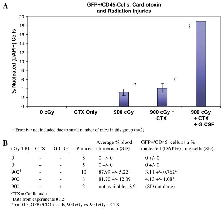
Methods: In separate experiments, mice received various doses of total-body irradiation followed by transplantation with whole bone marrow or various subpopulations of marrow cells (Lin(-/+), c-kit(-/+), Sca-1(-/+)) from GFP(+) (C57BL/6-TgN[ACTbEGFP]1Osb) mice. Some were given intramuscular cardiotoxin and/or mobilized with granulocyte colony-stimulating factor (G-CSF).

Results: The production of pulmonary epithelial cells from engrafted bone marrow was established utilizing green fluorescent protein (GFP) antibody labeling to rule out autofluorescence and deconvolution microscopy to establish the colocaliztion of GFP and cytokeratin and the absence of CD45 in lung samples after transplantation. More donor-derived lung cells (GFP(+)/CD45(-)) were seen with increasing doses of radiation (5.43% of all lung cells, 1200 cGy). In the 900-cGy group, 61.43% of GFP(+)/CD45(-) cells were also cytokeratin(+). Mobilization further increased GFP(+)/CD45(-) cells to 7.88% in radiation-injured mice. Up to 1.67% of lung cells were GFP(+)/CD45(-) in radiation-injured mice transplanted with Lin(-), c-kit(+), or Sca-1(+) marrow cells. Lin(+), c-kit(-), and Sca-1(-) subpopulations did not significantly engraft the lung.

Conclusions: We have established that marrow cells are capable of producing pulmonary epithelial cells and identified radiation dose and G-CSF mobilization as variables influencing the production of lung cells from marrow cells. Furthermore, the putative lung cell-producing marrow cell has the phenotype of a hematopoietic stem cell.


Figures

Figure 1
Figure 1
(A,B): GFP+ events are not due to autofluorescence. A: Lung samples from mice exposed to 900 cGy TBI were labeled with anti-GFP Alexafluor-594 (red) and DAPI nuclear counterstain (blue) and photographed using deconvolution fluorescence microscopy. There was red-green colocalization in all red or green cells observed in 10 random HPF from 5 samples (A: DAPI/FITC/rhodamine channels; B: FITC; C: rhodamine), indicating that all FITC+ cells were truly GFP+ . Corresponding images from a nonirradiated C57BL/6 mouse (D,E,F) and a C57BL/6 mouse exposed to 900 cGy TBI and transplanted with C57BL/6 WBM (G,H,I) indicate that no cells or structures appear green or label with the anti-GFP Ab. B: Cells from a lung sample from the 900-cGy group labeled with anti-GFP Alexafluor-594, photographed using deconvolution fluorescence microscopy, demonstrates clear red-green colocalization, indicating that green cells are truly GFP+ (A: FITC/rhodamine channels; B: FITC; C: rhodamine). A three-dimensional view of one cell from the deconvolved image (asterisk) confirms colocalization (D,E,F).
Figure 2
Figure 2
Radiation injury and the appearance of donor marrow–derived lung cells. (A): Samples from radiation-injured mice 1 month after TBI demonstrating increasing cellularity and thickening of the alveolar walls by H & E staining (A–D: light) and more GFP+ cells (E–H: FITC/rhodamine filters) with escalating doses of TBI. No radiation (A,E); 500 cGy (B,F); 900 cGy (C,G); 1200 cGy (D,H). (B,C): Results from the radiation injury experiment demonstrating that more donor marrow–derived lung cells (GFP+ and CD45) were seen in mice exposed to higher doses of radiation. (D): Samples from the radiation injury experiment were stained with anti-CD45 antibody using avidin Texas red as a secondary antibody and photographed using FITC (A), rhodamine (B), and FITC/rhodamine (C) filters. Native hematopoietic cells were considered to be CD45+ but GFP (asterisk). Donor-derived hematopoietic cells were considered to be GFP+ and CD45+ (small arrow). Donor-derived lung cells were considered to be GFP+ and CD45 (large arrow).
Figure 3
Figure 3
Donor-derived pulmonary epithelial cells.(A,B): Some of the donor-derived cells had the morphologic and phenotypic characteristics of pulmonary epithelial cells. Samples from all experiments were labeled with anti-CD45 (blue) and anti-cytokeratin (red) antibodies and photographed using deconvolution fluorescence microscopy. In A and B, the featured cells (arrow) are GFP+ (B: FITC filter), cytokeratin+ (C: rhodamine filter), and CD45 (D: DAPI filter), indicating that they represent donor-derived pulmonary epithelial cells (A: all filters). A three-dimensional view of the featured cells from the deconvolved image confirms colocalization of GFP and cytokeratin, but not CD45 (E,F,G).
Figure 4
Figure 4
Quantification of donor marrow–derived lung cells after radiation injury with G-CSF mobilization. (A,B): Results from the radiation injury with G-CSF mobilization experiment demonstrating that more donor marrow–derived lung cells (GFP+ and CD45) were seen in mobilized mice compared with nonmobilized mice.
Figure 5
Figure 5
Quantification of donor marrow–derived lung cells after radiation and cardiotoxin injuries. (A,B): Results from the radiation and cardiotoxin injuries experiment demonstrating that more donor marrow–derived lung cells (GFP+ and CD45) were seen in radiation and cardiotoxin–injured mice compared with radiation or cardiotoxin alone. When radiation and cardiotoxin–injured mice were mobilized with G-CSF, GFP+ /CD45 cells increased to 18.9% (2 mice).
Figure 6
Figure 6
Quantification of donor marrow–derived lung cells after radiation and cardiotoxin injuries, using different donor cell subpopulation. (A,B): Results from this experiment demonstrating higher levels of bone marrow engraftment and donor-derived lung cells (GFP+ and CD45) in radiation and cardiotox-in–injured mice transplanted with donor cells possessing the phenotype of hematopoietic stem cells (Sca-1+ , c-kit+ , Lin).

References

    1. Abedi M, Badiavas E, Lambert JF, Colvin G, Falanga V, Quesenberry PJ. Trafficking and transdifferentiation of bone marrow cells in a skin injury model. Exp Hematol. 2000;30:47.
    1. Ferrari G, Cusella-De Angelis G, Coletta M, et al. Muscle regeneration by bone marrow–derived myogenic progenitors. Science. 1998;279:1528–1530. - PubMed
    1. Gussoni E, Soneoka Y, Strickland CD, et al. Dystrophin expression in the mdx mouse restored by stem cell transplantation. Nature. 1999;401:390–394. - PubMed
    1. Hou Z, Nguyen Q, Frenkel B, et al. Osteoblast-specific gene expression after transplantation of marrow cells: implications for skeletal gene therapy. Proc Natl Acad Sci U S A. 1999;96:7294–7299. - PMC - PubMed
    1. Orlic D, Kajstura J, Chimenti S, et al. Bone marrow cells regenerate infarcted myocardium. Nature. 2001;410:701–705. - PubMed

Publication types

MeSH terms

Substances