Review
. 2006:68:279-305.
doi: 10.1146/annurev.physiol.68.040504.094635.

Brainstem circuits regulating gastric function

Affiliations
Review

Brainstem circuits regulating gastric function

R Alberto Travagli et al. Annu Rev Physiol. 2006.

Abstract

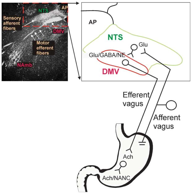
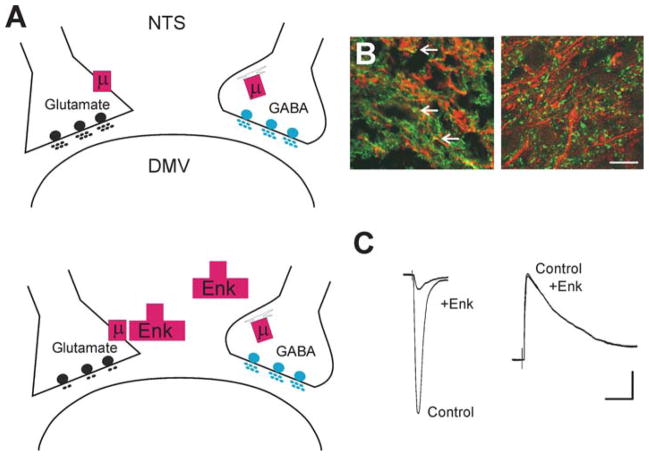
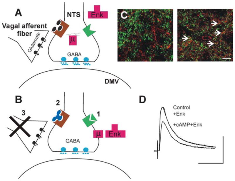
Brainstem parasympathetic circuits that modulate digestive functions of the stomach are comprised of afferent vagal fibers, neurons of the nucleus tractus solitarius (NTS), and the efferent fibers originating in the dorsal motor nucleus of the vagus (DMV). A large body of evidence has shown that neuronal communications between the NTS and the DMV are plastic and are regulated by the presence of a variety of neurotransmitters and circulating hormones as well as the presence, or absence, of afferent input to the NTS. These data suggest that descending central nervous system inputs as well as hormonal and afferent feedback resulting from the digestive process can powerfully regulate vago-vagal reflex sensitivity. This paper first reviews the essential "static" organization and function of vago-vagal gastric control neurocircuitry. We then present data on the opioidergic modulation of NTS connections with the DMV as an example of the "gating" of these reflexes, i.e., how neurotransmitters, hormones, and vagal afferent traffic can make an otherwise static autonomic reflex highly plastic.


Figures

Figure 1
Figure 1
Cytoarchitecture of the dorsal vagal complex. Darkfield photomicrograph of a coronal section of rat brainstem at the level of the area postrema/intermediate level following application of HRP crystals to the subdiaphragmatic vagus. Note the intense labeling of the nucleus tractus solitarius (NTS), dorsal motor nucleus of the vagus (DMV), and nucleus ambiguus (NAmb). An area of the photomicrograph (dotted line) has been expanded into cartoon form to allow a more detailed illustration of the brainstem circuitry of gastrointestinal (GI) vago-vagal reflexes. Note that vagal afferent neurons, whose cell bodies lie in the nodose ganglion, receive sensations from the GI tract. The central terminals of these afferent fibers enter the brainstem via the tractus solitarius and terminate within the NTS, utilizing principally glutamate as their neurotransmitter. These afferent signals are integrated by neurons of the NTS that project to, among other areas, the adjacent DMV using, mainly, glutamate, GABA, or NE as neurotransmitters. Neurons of the DMV are the preganglionic parasympathetic motoneurons that provide the motor output to the GI tract via the efferent vagus, where they release acetylcholine onto their postganglionic target. Postganglionic parasympathetic neurons are either excitatory [cholinergic (ACh)] or inhibitory [nonadrenergic, noncholinergic (NANC)].
Figure 2
Figure 2
Differential modulation of synaptic transmission. Depiction of the synaptic connections between the NTS and the DMV. (A) Under control conditions, the μ-opioid receptor (μ) is expressed on the nerve terminal of glutamatergic NTS profiles apposing DMV neurons; by contrast, on GABAergic nerve terminals, the μ-opioid receptor is internalized and associated with the Golgi apparatus (upper panel). Opioid agonists (e.g., Enk) can act to inhibit glutamate synaptic transmission, but not GABAergic transmission (lower panel). (B) High-power photomicrographs of the cloned μ-opioid receptor (MOR1)-immunoreactivity (-IR; TRITC filters, red) and γ-glutamyl glutamate-IR (Glu-IR; FITC filters, green; left panel) used as a marker for glutamate nerve terminals, and glutamic acid decarboxylase-IR (GAD-IR; FITC filters, green; right panel) used as a marker for GABAergic nerve terminals in the rat DVC. Note that MOR1-IR is colocalized with glutamate nerve terminals (yellow; arrows) but not with GABA nerve terminals. Scale bar: 15 μm. (C) Representative traces of evoked excitatory postsynaptic currents (eEPSCs) (left panel) and evoked inhibitory postsynaptic currents (eIPSCs) (right panel) in gastric-projecting DMV neurons voltage-clamped at −60 mV and −50 mV, respectively, evoked by electrical stimulation of the NTS. Perfusion with Enk inhibits the eEPSCs but not eIPSCs. Scale bar: 50 pA and 30 ms.
Figure 3
Figure 3
Increasing the levels of cAMP in the brainstem induces receptor trafficking in NTS nerve terminals. (A) In control conditions, i.e., when the levels of cAMP are low either because the tonic release of glutamate from vagal afferent fibers activates group II metabotropic glutamate receptors (left) or because Gαpled receptors are not activated (right), the terminals of GABAergic neurons in the NTS store μ-opioid receptors (μ) in internal compartments associated with the Golgi complex. In this situation, μ-opioid agonists (e.g., Enk) cannot modulate the release of GABA onto DMV neurons. (B) Following increases in cAMP levels within the GABAergic nerve terminal, for example, by (1) activation of a receptor coupled to Gα., TRH or CCK, (2) antagonism of group II metabotropic glutamate receptors, or (3) removal of tonic vagal afferent input, μ-opioid receptors are released from the Golgi apparatus and translocated to the nerve terminal membrane, where opioid agonists can inhibit GABA synaptic transmission between the NTS and the DMV. (C) High-power photomicrographs of the cloned μ-opioid receptor (MOR1)-immunoreactivity (-IR; TRITC filters, red) and glutamic acid decarboxylase-IR (GAD-IR; FITC filters, green) used as a marker for GABAergic nerve terminals in the rat DVC. In control conditions (left panel), note the absence of MOR- and GAD-IR colocalized profiles. Following vagal afferent rhizotomy (right panel), many nerve terminals show MOR- and GAD-IR colocalized profiles (yellow; arrows). Images represent three-dimensional reconstructions from Z-stack image series. Scale bar: 10 μm. (D) Representative traces of eIPSCs evoked in a gastric-projecting DMV neuron voltage-clamped at −50 mV. Perfusion with Enk does not affect the amplitude of IPSCs evoked by electrical stimulation of the NTS. Following five minutes’ perfusion with substances that increase the cAMP levels, however, reapplication of Enk reduces the amplitude of the eIPSCs. Scale bar: 50 ms and 200 pA.

References

    1. Wood JD. Physiology of the enteric nervous system. In: Johnson LR, editor. Physiology of the Gastrointestinal Tract. New York: Raven; 1987. pp. 67–110.
    1. Goyal RK, Hirano I. The enteric nervous system. N Engl J Med. 1996;334:1106–15. - PubMed
    1. Costa M, Brookes SJH, Hennig GW. Anatomy and physiology of the enteric nervous system. Gut. 2000;47:15–19. - PMC - PubMed
    1. Bornstein JC, Costa M, Grider JR. Enteric motor and interneuronal circuits controlling motility. Neurogastroenterol Motil. 2004;16(Suppl 1):34–38. - PubMed
    1. Goyal RK, Paterson WG. Esophageal motility. In: Wood JD, editor. Handbook of Physiology—The Gastrointestinal System I. Bethesda, MD: Am. Phys. Soc; 1989. pp. 865–908.

Publication types

LinkOut - more resources