

The structural basis of arrestin-mediated regulation of G-protein-coupled receptors
- PMID: 16460808
- PMCID: PMC2562282
- DOI: 10.1016/j.pharmthera.2005.09.008
The structural basis of arrestin-mediated regulation of G-protein-coupled receptors
Abstract
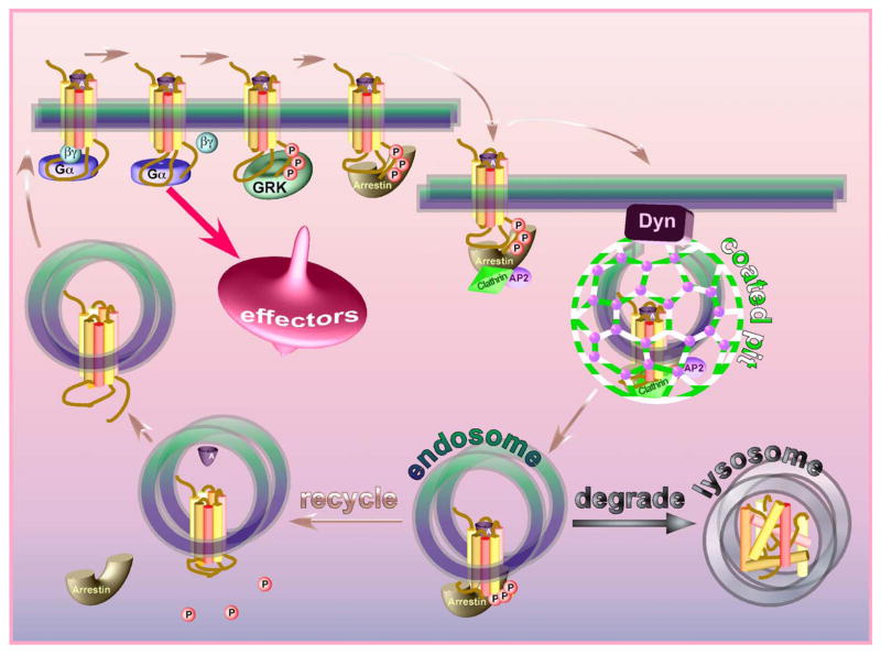
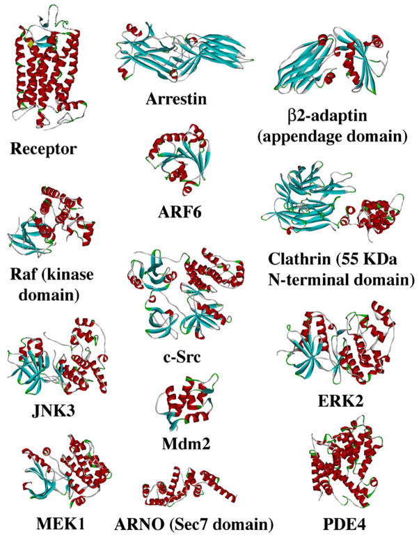
The 4 mammalian arrestins serve as almost universal regulators of the largest known family of signaling proteins, G-protein-coupled receptors (GPCRs). Arrestins terminate receptor interactions with G proteins, redirect the signaling to a variety of alternative pathways, and orchestrate receptor internalization and subsequent intracellular trafficking. The elucidation of the structural basis and fine molecular mechanisms of the arrestin-receptor interaction paved the way to the targeted manipulation of this interaction from both sides to produce very stable or extremely transient complexes that helped to understand the regulation of many biologically important processes initiated by active GPCRs. The elucidation of the structural basis of arrestin interactions with numerous non-receptor-binding partners is long overdue. It will allow the construction of fully functional arrestins in which the ability to interact with individual partners is specifically disrupted or enhanced by targeted mutagenesis. These "custom-designed" arrestin mutants will be valuable tools in defining the role of various interactions in the intricate interplay of multiple signaling pathways in the living cell. The identification of arrestin-binding sites for various signaling molecules will also set the stage for designing molecular tools for therapeutic intervention that may prove useful in numerous disorders associated with congenital or acquired disregulation of GPCR signaling.
Figures
References
-
- Altenbach C, Cai K, Khorana HG, Hubbell WL. Structural features and light-dependent changes in the sequence 306–322 extending from helix VII to the palmitoylation sites in rhodopsin: a site-directed spin-labeling study. Biochemistry. 1999a;38:7931–7937. - PubMed
-
- Altenbach C, Klein-Seetharaman J, Hwa J, Khorana HG, Hubbell WL. Structural features and light-dependent changes in the sequence 59–75 connecting helices I and II in rhodopsin: a site-directed spin-labeling study. Biochemistry. 1999b;38:7945–7949. - PubMed
-
- Altenbach C, Cai K, Klein-Seetharaman J, Khorana HG, Hubbell WL. Structure and function in rhodopsin: mapping light-dependent changes in distance between residue 65 in helix TM1 and residues in the sequence 306–319 at the cytoplasmic end of helix TM7 and in helix H8. Biochemistry. 2001a;40:15483–15492. - PubMed
-
- Altenbach C, Klein-Seetharaman J, Cai K, Khorana HG, Hubbell WL. Structure and function in rhodopsin: mapping light-dependent changes in distance between residue 316 in helix 8 and residues in the sequence 60–75, covering the cytoplasmic end of helices TM1 and TM2 and their connection loop CL1. Biochemistry. 2001b;40:15493–15500. - PubMed
-
- Angers S, Salahpour A, Bouvier M. Dimerization: an emerging concept for G protein-coupled receptor ontogeny and function. Annu Rev Pharmacol Toxicol. 2002;42:409–435. - PubMed
Publication types
MeSH terms
Substances
Grants and funding
LinkOut - more resources
Full Text Sources
Other Literature Sources
Molecular Biology Databases
