Comparative Study
. 2006 Apr;13(4):756-65.
doi: 10.1016/j.ymthe.2005.12.008. Epub 2006 Feb 7.

Evaluation of adenovirus vectors containing serotype 35 fibers for vaccination

Affiliations
Comparative Study

Evaluation of adenovirus vectors containing serotype 35 fibers for vaccination

Nelson DiPaolo et al. Mol Ther. 2006 Apr.

Abstract

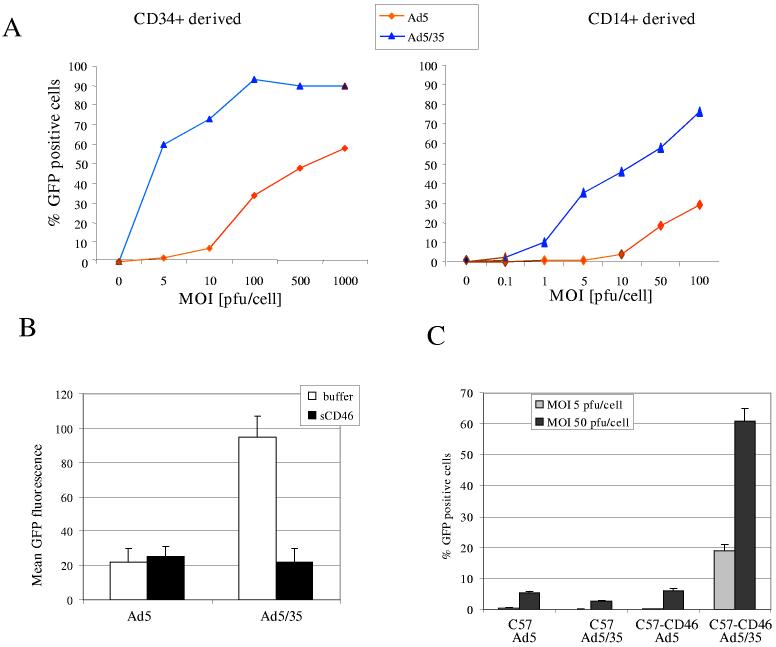
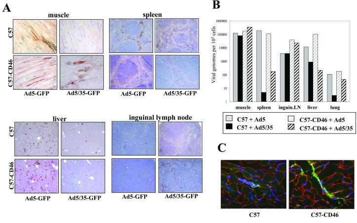
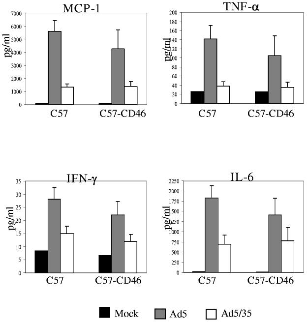
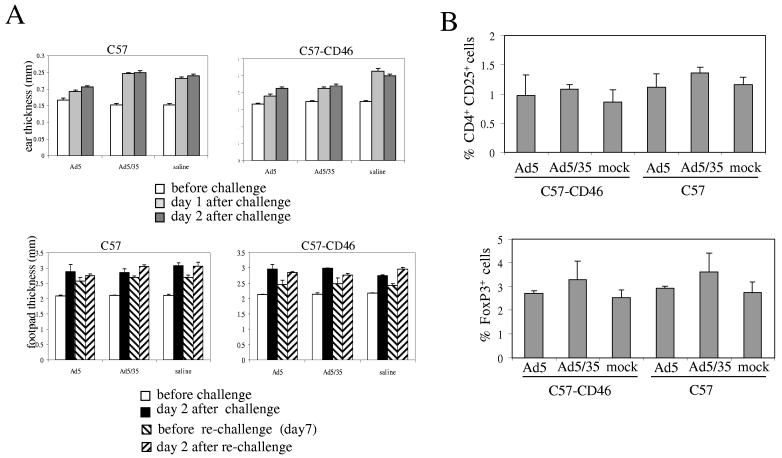
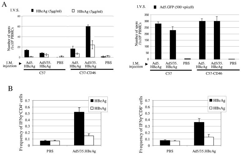
In contrast to commonly used serotype 5-based adenovirus (Ad) vectors, Ad's containing fibers derived from B-group serotype 35 (Ad5/35) efficiently transduce human DCs ex vivo and appear to target antigen-presenting cells after intravenous injection into baboons. Based on this, Ad5/35 vectors could be valuable tools for immunotherapy and vaccination. On the other hand, a number of studies indicate that signaling through the B-group Ad receptor, CD46, can cause tolerance or immunosuppression. Since mice do not express CD46 in a human-like pattern, we studied the in vivo properties of Ad5/35 in transgenic mice that express CD46 in a pattern and at a level similar to those of humans. Hypersensitivity assays and analyses of frequencies of regulatory T cells and T cell responses did not indicate that Ad5/35 injection exerts detrimental effects on the host's immune system. An Ad5/35 vector expressing a model antigen was able to trigger a strong T cell response against the test antigen after intramuscular injection. Overall, compared to Ad5 vectors, Ad5/35 vectors had a better safety profile, reflected by lower serum levels of proinflammatory cytokines.


Figures

Figure 1
Figure 1
Assessment of Ad5/35 vectors for transduction of dendritic cells. A) Transduction of human dendritic cells (DC) with first-generation Ad5 and Ad5/35 vectors containing a CMV-GFP expression cassette. Immature human DCs (>95% positive for CD11c) derived either from CD34+ cells (left panel) or peripheral CD14+ monocytes (right panel) were infected at different MOIs for 3 hours. GFP expression was analyzed by flow cytometry 24 hours post-infection. B) Transduction of DCs in the presence of soluble CD46. CD34+ derived DCs were transduced with Ad5-GFP or Ad5/35-GFP at an MOI of 10pfu/cell in the presence of an excess of soluble CD46 (as a competitor) and GFP expression was analyzed 24 hours later by flow cytometry. C) Transduction of DC from CD46 transgenic mice with Ad vectors. Myeloid DC from C57 and C57-CD46 mice were infected with first-generation Ad5 and Ad5/35 vectors at an MOI of 5 and 50 pfu/cell. The percentage of GFP expressing cells was analyzed 24 hours after infection by flow cytometry.
Figure 2
Figure 2
Biodistribution of Ad mediated transgene expression and vector genomes in C57 and C57-CD46 mice after intramuscular injection of Ad5.GFP and Ad5/35.GFP. A) GFP expression in organ sections 72 hours after intramuscular injection of 5×10⁹ pfu of Ad5.GFP and Ad5/35.GFP. Tissue sections were analyzed for GFP expression by immunohistochemistry with anti-GFP antibodies. B) Quantitative comparison of viral genomes present in major organs at 72 hours after intramuscular Ad injection. The genome concentration was expressed as the number of viral genomes per 107 cells (assuming that the mass of a diploid human genome is 6pg). qPCR results for vector genomes were equalized based on qPCR data for an endogenous (two copies/genome) mouse GAPDH gene. The average of 3 independent tissue samples is shown. Standard deviation was less than 10%C) Detection of Ad5/35 particles around the injection site. Mice were injected IM with 5×10⁹pfu Ad5/35.GFP in 40μl of PBS. One hour after injection, mice were sacrificed. Sections (8μm) of muscle tissue around the injection site were analyzed by immunofluorescence. Viral particles were detected using anti-Ad5 hexon-FITC antibodies (green). Muscle cells were stained with anti-laminin antibodies (red). Cell nuclei are blue.
Figure 3
Figure 3
Innate immune responses upon IM injection of Ad5/35 into mice. Plasma levels of MCP-1, TNF-α, IFN-γ, and IL-6 at 6 hours after IM injection of Ad vectors. Mice were injected with PBS (Mock) or with 5×10⁹ pfu of Ad5.GFP or Ad5/35.GFP. Plasma samples from three individual mice per virus were collected and analyzed in duplicate by cytometric bead array for cytokine and chemokine levels.
Figure 4
Figure 4
Absence of immunosuppression upon Ad5/35 infectionA) CD4- and CD8 mediated hypersensitivity reactions. C57 or CD57-CD46 mice were injected intramuscularly with saline, 5×10⁹pfu of Ad5.GFP or Ad5/35.GFP. Upper panel: Contact hypersensitivity: Six hours after virus injection mice were sensitized with dinitrofluorobenzene. Six day later, mice were challenged and the ear thickness was measured before challenge and 24 and 48 hours after challenge. Lower Panel: Delayed type hypersensitivity: Six hours after virus injection, mice were sensitized with KLH. Six day later, mice were challenged and the food pad thickness was measured before challenge and 48 hours after challenge. To assess potential long-term effects of Ad injection, mice were re-challenged with KLH and foot pad thickness was measured before and after re-challenging. (N=3)B) Analysis of regulatory T-cells. C57 or CD57-CD46 mice were injected with 5×10⁹pfu of Ad5.GFP or Ad5/35.GFP. Seven days later splenocytes and lymph nodes were harvested and analyzed by flow cytometry for the percentage of CD4+CD25+ and FoxP3+ cells (N=3).
Figure 5
Figure 5
Analysis of frequencies of IFNγ-producing T-cells specific to a model antigen or adenovirus vector.A) ELIsport analyses of IFNγ-producing T-cells frequencies. C57 or C57-CD46 mice were injected intramuscularly with 5×10⁹ pfu of Ad5.HBeAg, Ad5/35.HBeAg, or PBS. Twelve days later, spleen cells of naïve syngeneic animals were obtained and pulsed with 5μg/ml of recombinant HBeAg and HBsAg (as a negative control) (left panel) or transduced with 25pfu (=500 viral particles) of Ad.5GFP (right panel). On day 14, vaccinated animals were sacrificed, splenocytes were collected and 1×10⁶ cells were mixed with 1×10⁶ ex vivo pulsed splenocytes for in vitro sensitization. After 24 hours of incubation in 96 well plates, cells were plated in anti-IFNγ-coated wells of an ELIspot plate. Twenty-four hours later, plates were washed and the spots of IFNγ producing T-cells were counted. The number of spots was expressed as the average +/− the standard deviation. N=3 animals per group.B) Frequency of IFNγ+ CD4+ cells (left panel) and IFNγ+ CD8+ cells (right panel) analyzed by intracellular cytokine staining. C57-CD46 mice were injected intramuscularly with 5×10⁹ pfu of Ad5/35.HBeAg, or PBS. Twelve days after immunization, splenocytes were harvested and cultured with recombinant HBeAg and (control) HBsAg for 5 hours in the presence of Brefeldin. Cells were then stained for CD4 and CD8 and intracellular IFNγ. Shown is the percentage of IFNγ+CD4+ in all CD4+ cells and IFNγ+CD8+ in all CD8+ cells. (N=3)
Figure 6
Figure 6
Analysis of memory T-cells against Ad vectors in human PBMCs by ELIspot for IFNγ expressing T-cells.PBMCs were obtained from normal donors that have never done research with Ad vectors, from normal donors that worked with Ad vectors, and from Senegalese, stage III breast cancer patients (before chemotherapy). Cells were transduced with the indicated Ad vectors at an MOI of 200 viral particles/cell and the number of IFNγ expressing T-cells was analyzed by ELIspot 24 hours later. N=3
Figure 7
Figure 7
Cross-reactivity of anti-fiber knob T-cells.A) Memory T-cells against fiber knob domains. PBMC from two normal donors that are involved in adenovirus research were incubated with 0.2μg/ml of recombinant Ad5 or Ad35 fiber knob domains (produced in E.coli) or control peptide (HBeAg) and the frequency of IFNγ expressing T-cells was analyzed by ELIspot 24 hours later. (N=3) B) Crossreactivity of T-cells against fiber knobs. T-cells were primed by incubating PBMC from a healthy donor with recombinant Ad5 or Ad35 fiber knob. Subsequently T-cells were incubated for 7 days in the presence of IL-2 and then boosted with dendritic cells pulsed with control peptide (Co), Ad5, or Ad35 fiber knob. The frequency of IFNγ expressing T-cells was analyzed by ELIspot 24 hours later. (N=3)

References

    1. Tatsis N, Ertl HC. Adenoviruses as vaccine vectors. Mol Ther. 2004;10:616–629. - PMC - PubMed
    1. Cohen J. AIDS research. Merck reemerges with a bold AIDS vaccine effort. Science. 2001;292:24–25. - PubMed
    1. Muruve DA, Barnes MJ, Stillman IE, Libermann TA. Adenoviral gene therapy leads to rapid induction of multiple chemokines and acute neutrophil-dependent hepatic injury in vivo. Hum Gene Ther. 1999;10:965–976. - PubMed
    1. Rea D, et al. Highly efficient transduction of human monocyte-derived dendritic cells with subgroup B fiber-modified adenovirus vectors enhances transgene-encoded antigen presentation to cytotoxic T cells. J Immunol. 2001;166:5236–5244. - PubMed
    1. Ni S, et al. Evaluation of biodistribution and safety of adenovirus vectors containing group B fibers after intravenous injection into baboons. Hum Gene Ther. 2005;16:664–677. - PMC - PubMed

Publication types

MeSH terms