Review
. 2006 Feb;35(4):238-49.
doi: 10.1016/j.semarthrit.2005.08.002.

Description of 12 cases of nephrogenic fibrosing dermopathy and review of the literature

Affiliations
Review

Description of 12 cases of nephrogenic fibrosing dermopathy and review of the literature

Fabian A Mendoza et al. Semin Arthritis Rheum. 2006 Feb.

Abstract

Objectives: To review the clinical and laboratory features of 12 cases of nephrogenic fibrosing dermopathy (NFD) studied at our institution and of 70 previously described cases in the literature.

Methods: Clinical evaluation and laboratory studies of 12 patients with NFD associated with chronic hemodialysis or peritoneal dialysis for end-stage renal disease and a review of 23 previous publications describing 70 patients with this disease.

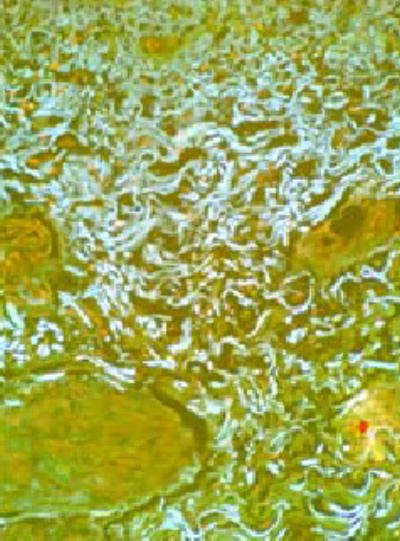
Results: Eleven patients undergoing chronic hemodialysis and 1 patient undergoing chronic peritoneal dialysis for end-stage renal failure developed a severe and progressive cutaneous fibrotic process with woody induration of legs, thighs, hands, and forearms, and severe loss of motion and flexion contractures in multiple joints. Several patients displayed systemic involvement including fibrosis of muscles, myocardium, and lungs and marked elevations of the erythrocyte sedimentation rate and/or C-reactive protein. Three patients died within 2 years of symptom onset. A review of previously published reports of this disorder confirmed the presence of systemic involvement and a poor prognosis with a high mortality rate.

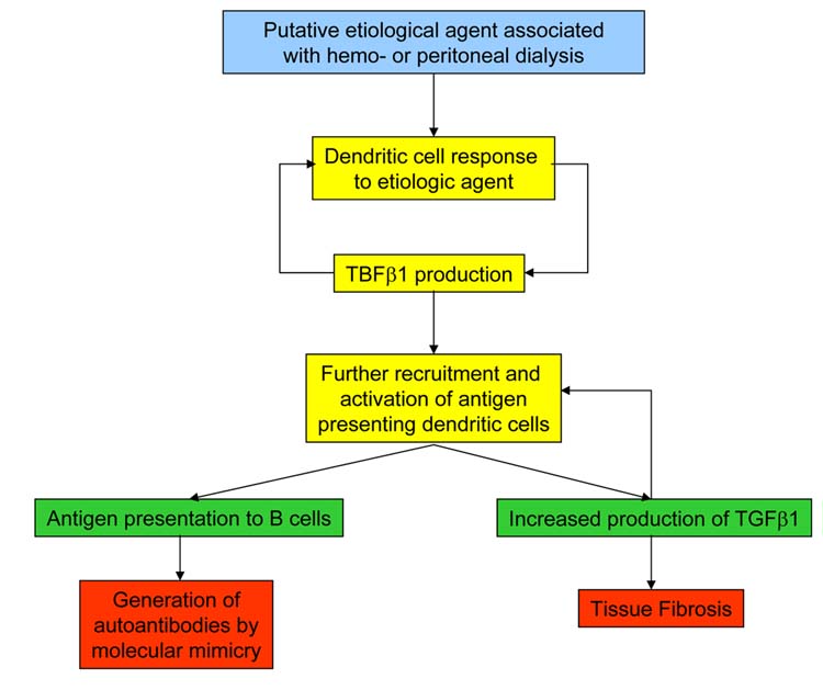
Conclusions: NFD is a severe and usually progressive systemic fibrotic disease affecting the dermis, subcutaneous fascia, and striated muscles. It also appears that the disease can cause fibrosis of lungs, myocardium, and other organs.


Figures

Figure 1
Figure 1. Cutaneous involvement
Note marked skin thickening including sclerodactyly and involvement of the dorsum of the hands (A, B) and “peau de orange” appearance in several affected areas (C, D). Severe fixed flexion contractures in fingers and wrist joints are apparent (C). Involvement of the lower legs (E) and a marked hypertrophic and neovascularized scar at a site of a minor dermal injury are illustrated (F).
Figure 2
Figure 2
Diagram illustrating the pattern of cutaneous involvement in the patients of the cohort we studied.
Figure 3
Figure 3. Histopathology of skin and subcutaneous tissues
(A) Hematoxylineosin and (B) trichrome stains. Note marked thickening of the entire dermis and subcutaneous tissues with severe accumulation of thick collagen bundles separated by numerous clefts. (C) Hematoxylineosin showing thick tracts of fibrous tissue extending into the adipose layer resulting in microlobular architecture and in thickening of the fascia. Magnification 60X.
Figure 4
Figure 4. Morphology of dermal and subdermal collagen fibers
Thick and tortuous collagen bundles in the deep dermis and subcutaneous tissue of affected skin viewed under fluorescence microscopy with a triple-band pass filter. Magnification 400X.
Figure 5
Figure 5. Co-expression of CD68 and FXIIIa in infiltrating cells in skin
Cells were dual-stained for CD68 (red) and FXIIIa (green). Both antibodies were diluted 1:100 and simultaneously applied to the section for 40 min. Unbound antibodies were removed with 3 washes of 2 min each of PBS. The secondary antibodies; rabbit-anti-mouse Cy3 (1:100) and donkey-anti-goat-FITC (1:100) were applied to the section for 40 min and the unbound proteins washed off. Sections were counterstained with DapI and viewed with an epi-fluorescence microscope. Panel A shows two cells that are positive for FXIIIa. Panel B: the same cells are viewed through the red filter to detect CD68. Panel C: the cells are viewed through the triple bandpass filter to detect the co-expression of FXIIIa and CD68. The cells appear yellow when there is co-localization of the two antibodies. Magnitication 400X.
Figure 6
Figure 6. Histopathology of muscle
Trichrome stain. Note remarkable accumulation of fibrotic tissue in the interfascicular septae and surrounding muscle fibers. Magnification 200X.
Figure 7
Figure 7. Histopathology of myocardium
Trichrome stain. Note thickening of pericardium and accumulation of large amounts of fibrous tissue surrounding and between myocardial fibers. Magnification 200X.
Figure 8
Figure 8. Histopathology of lung tissue
Trichrome stain. (A) Lung parenchyma showing interstitial fibrosis with severe distortion of alveolar architecture and moderate inflammatory cell infiltrate. (B) Small pulmonary arterioles showing marked thickening and fibrosis of adventitial layer. Interstitial fibrosis is also observed. Magnification 100X.
Figure 9
Figure 9. Expression of TGFβ in NFD skin
Fixed skin slides were stained with a fluorescein-labeled TGFβ specific antibody (green). The TGFβ antibody was diluted 1:100 and applied to the section for 40 min. The unbound antibody was removed with 3 washes of 2 min each of PBS. The secondary antibody was donkey-anti-goat-FITC (1:100) and it was applied to the section for 40 min and the unbound proteins washed off. Sections were counterstained with DapI and viewed with an epi-fluorescence microscope. Note the abundant expression of TGFβ epitopes throughout the dermis in dermal fibroblasts and infiltrating inflammatory cells.
Figure 10
Figure 10
Schematic diagram of NFD pathogenesis through the involvement of dendritic cells and TGFβ.
Figure 11
Figure 11
Schematic diagram of NFD pathogenesis through the involvement of circulating fibrocytes.

Comment in

References

    1. Cowper SE, Robin HS, Steinberg SM, Su LD, Gupta S, LeBoit PE. Scleromyxoedema-like cutaneous disease in renal-dialysis patients. Lancet. 2000;356:1000–01. - PubMed
    1. Cowper SE, Su LD, Bhawan J, Robin HS, LeBoit PE. Nephrogenic fibrosing dermopathy. Am J Dermatopathol. 2001;23:383–93. - PubMed
    1. http://www.icnfdr.org/ Accessed June 9, 2005.
    1. Ting WW, Stone MS, Madison KC, Kurtz K. Nephrogenic fibrosing dermopathy with systemic involvement. Arch Dermatol. 2003;139:903–06. - PubMed
    1. Jimenez SA, Arlett CM, Sandorfi N, Derk C, Latinis K, Sawaya H, et al. Dialysis-Associated Systemic Fibrosis (Nephrogenic Fibrosing Dermopathy) Arthritis Rheum. 2004;50:2660–66. - PubMed