. 2006 Feb 20;203(2):413-24.
doi: 10.1084/jem.20051720. Epub 2006 Feb 6.

Yellow fever vaccine YF-17D activates multiple dendritic cell subsets via TLR2, 7, 8, and 9 to stimulate polyvalent immunity

Affiliations

Yellow fever vaccine YF-17D activates multiple dendritic cell subsets via TLR2, 7, 8, and 9 to stimulate polyvalent immunity

Troy Querec et al. J Exp Med. .

Abstract

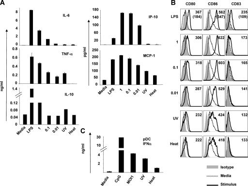
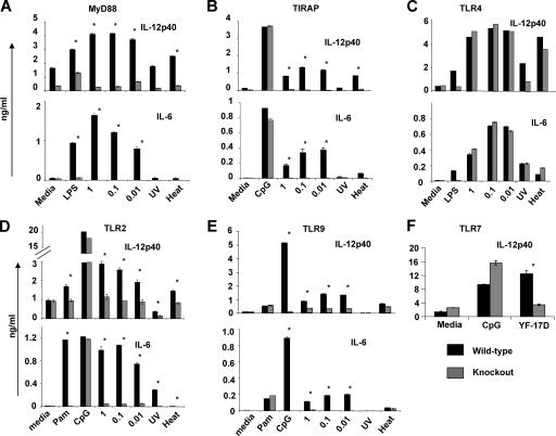
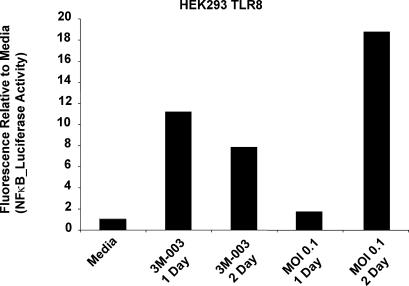
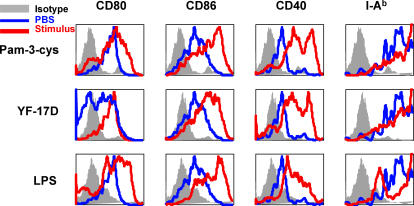
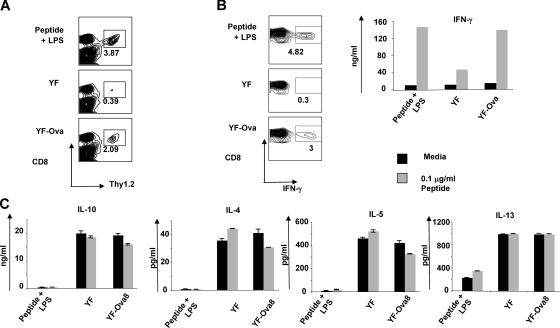
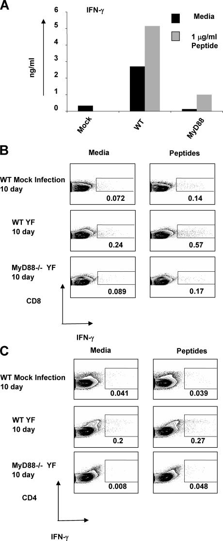
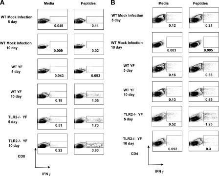
The live attenuated yellow fever vaccine 17D (YF-17D) is one of the most effective vaccines available, with a 65-yr history of use in >400 million people globally. Despite this efficacy, there is presently no information about the immunological mechanisms by which YF-17D acts. Here, we present data that suggest that YF-17D activates multiple Toll-like receptors (TLRs) on dendritic cells (DCs) to elicit a broad spectrum of innate and adaptive immune responses. Specifically, YF-17D activates multiple DC subsets via TLRs 2, 7, 8, and 9 to elicit the proinflammatory cytokines interleukin (IL)-12p40, IL-6, and interferon-alpha. Interestingly, the resulting adaptive immune responses are characterized by a mixed T helper cell (Th)1/Th2 cytokine profile and antigen-specific CD8+ T cells. Furthermore, distinct TLRs appear to differentially control the Th1/Th2 balance; thus, whilst MyD88-deficient mice show a profound impairment of Th1 cytokines, TLR2-deficient mice show greatly enhanced Th1 and Tc1 responses to YF-17D. Together, these data enhance our understanding of the molecular mechanism of action of YF-17D, and highlight the potential of vaccination strategies that use combinations of different TLR ligands to stimulate polyvalent immune responses.


Figures

Figure 1.
Figure 1.
YF-17D activates human monocyte–derived DCs and plasmacytoid DCs. Human mDCs were cultured with LPS; YF-17D at an MOI of 1, 0.1, or 0.01; or they were YF-17D UV or heat inactivated. Cytokines (A) and costimulatory molecule expression (B) were measured after 48 h. (A) The mean fluorescent intensities, are indicated at the top left of each histogram. (B, top) In the histograms, the numbers in parentheses represent the mean fluorescent intensities of the DCs cultured in media alone. (C) PDCs were isolated from human blood using BDCA-2 microbeads and culture for 48 h before measuring IFN-α. Error bars in A and C represent standard deviation. These data are representative of at least four independent experiments.
Figure 2.
Figure 2.
YF-17D activates DCs through multiple TLRs and multiple T LR adaptor proteins. CD11c+ DCs were isolated from the spleens of Flt-3L–injected knockout and age-matched, sex-matched, wild-type mice and cultured for 48 h in the presence of TLR agonists and YF. Cell culture supernatants were assayed for IL-12p40 and IL-6. (A) MyD88−/− mice versus control mice; (B) TIRAP−/− mice versus control mice; (C) TLR4−/− mice versus control mice; (D) TLR2−/− mice versus control mice; (E) TLR9−/− mice versus control mice; and (F) TLR7−/− mice versus control mice. Error bars represent standard deviation. These data are representative of at least four independent experiments. *, indicates statistically significant difference (P < 0.005 by paired Student's t test) between wild type (black) and knockout (gray) DCs.
Figure 3.
Figure 3.
YF-17D signals through TLR 8. Stably transfected HEK293 cells expressing human TLR8 and an NFκB-inducible luciferase reporter were stimulated with the TLR7/8 agonist 3M-003 or an MOI of 0.1 of YF-17D. After 1 and 2 d, the cells were lysed an analyzed for luciferase activity.
Figure 4.
Figure 4.
YF-17D induces DC activation in vivo. Mice were injected with YF-17D, LPS, Pam3Cys, or PBS subcutaneously and the draining lymph nodes were collected 24 h later. Costimulatory molecule expression on CD11c+ DCs was analyzed by flow cytometry.
Figure 5.
Figure 5.
YF-17D induces efficient induction of antigen-specific CD8+ T cells, and a mixed Th1/Th2 cytokine profile in vivo. Mice were injected with 2.5 × 10⁶ OT-I TCR transgenic T cells and allowed to rest overnight. YF-Ova8 vaccinations were administered subcutaneously and the draining lymph nodes were collected 5 d later. A portion of cells was stained directly ex vivo for CD8 and Thy 1.2 to evaluate clonal expansion of SIINFEKL-specific CD8+ T cells by FACS (A). The remaining cells were placed into culture and restimulated with SIINFEKL or cultured in media alone. After 3 d, the induction of IFN-γ, IL-4, IL-5, and IL-13 cytokines was measured by intracellular FACS staining (B, IFN-γ) or by ELISA (B and C). Error bars represent standard deviation. These data are representative of three independent experiments.
Figure 6.
Figure 6.
Induction of Th1 and Tc1 responses by YF-17D is dependent on MyD88. Wild-type or MyD88−/− mice were vaccinated subcutaneously with YF-17D. Control mice were injected with supernatants from mock transfected SW-480 cells. 10 d later, the draining lymph nodes were harvested and single cell suspensions were cultured in vitro in media alone or with a combination of CD8+ T and CD4+ T cell epitopes from YF-17D as described in Materials and methods. 3 d later, the induction of IFN-γ was measured by ELISA (A) and by intracellular cytokine staining (B and C). These data are representative of three independent experiments with three mice per group in each experiment.
Figure 7.
Figure 7.
TLR2 exerts an inhibitory effect on Th1 and Tc1 responses against YF-17D. Wild-type or TLR2−/− mice were vaccinated subcutaneously with YF-17D. Control mice were injected with supernatants from mock transfected SW-480 cells. 5 or 10 d later, the draining lymph nodes were harvested and single cell suspensions cultured in vitro in media alone, or with a combination of CD8+ T and CD4+ T cell epitopes from YF-17D. 3 d later, the induction of IFN-γ was measured by intracellular cytokine staining (A and B). These data are representative of four independent experiments with three mice per group in each experiment.

References

    1. Theiler, M., and H.H. Smith. 1937. The use of yellow fever virus modified by in vitro cultivation for human immunization. J. Exp. Med. 65:787–800. - PMC - PubMed
    1. Theiler, M., and H.H. Smith. 1937. The effect of prolonged cultivation in vitro upon the pathogenicity of yellow fever virus. J. Exp. Med. 65:767–786. - PMC - PubMed
    1. Niedrig, M., M. Lademann, P. Emmerich, and M. Lafrenz. 1999. Assessment of IgG antibodies against yellow fever virus after vaccination with 17D by different assays: neutralization test, haemagglutination inhibition test, immunofluorescence assay and ELISA. Trop. Med. Int. Health. 4:867–871. - PubMed
    1. Poland, J.D., C.H. Calisher, T.P. Monath, W.G. Downs, and K. Murphy. 1981. Persistence of neutralizing antibody 30-35 years after immunization with 17D yellow fever vaccine. Bull. World Health Organ. 59:895–900. - PMC - PubMed
    1. Co, M.D., M. Terajima, J. Cruz, F.A. Ennis, and A.L. Rothman. 2002. Human cytotoxic T lymphocyte responses to live attenuated 17D yellow fever vaccine: identification of HLA-B35-restricted CTL epitopes on nonstructural proteins NS1, NS2b, NS3, and the structural protein E. Virology. 293:151–163. - PubMed

Publication types

MeSH terms