. 2006 Feb 20;203(2):297-303.
doi: 10.1084/jem.20051923. Epub 2006 Feb 6.

ATM regulates ATR chromatin loading in response to DNA double-strand breaks

Affiliations

ATM regulates ATR chromatin loading in response to DNA double-strand breaks

Myriam Cuadrado et al. J Exp Med. .

Abstract

DNA double-strand breaks (DSBs) are among the most deleterious lesions that can challenge genomic integrity. Concomitant to the repair of the breaks, a rapid signaling cascade must be coordinated at the lesion site that leads to the activation of cell cycle checkpoints and/or apoptosis. In this context, ataxia telangiectasia mutated (ATM) and ATM and Rad-3-related (ATR) protein kinases are the earliest signaling molecules that are known to initiate the transduction cascade at damage sites. The current model places ATM and ATR in separate molecular routes that orchestrate distinct pathways of the checkpoint responses. Whereas ATM signals DSBs arising from ionizing radiation (IR) through a Chk2-dependent pathway, ATR is activated in a variety of replication-linked DSBs and leads to activation of the checkpoints in a Chk1 kinase-dependent manner. However, activation of the G2/M checkpoint in response to IR escapes this accepted paradigm because it is dependent on both ATM and ATR but independent of Chk2. Our data provides an explanation for this observation and places ATM activity upstream of ATR recruitment to IR-damaged chromatin. These data provide experimental evidence of an active cross talk between ATM and ATR signaling pathways in response to DNA damage.


Figures
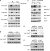
Figure 1.
Figure 1.
Analysis of ATM, Chk2, and Chk1 activation in response to DNA damage. (A) Western blot detection of ATM-1981P, ATM, Chk2-T68P, Chk2, Chk1-S345P, and Chk1 in Raji human lymphoblastoid cells exposed to DNA-damaging conditions. Where indicated, ATM activity was inhibited by a previous 15-min treatment with 20 μM wortmannin (WM). (B) Evaluation of Chk1 phosphorylation dynamics in control and AT human cells in response to DNA damage. (C) Western blot analysis of ATR, Chk1-S345P, Chk1, ATM-1981P, and ATM levels in extracts from control (ATRwt) and ATR hypomorphic (ATRSeckel) human cells irradiated with 20 Gy (or mock irradiated [C]). β-actin levels were evaluated as a loading control. (D) Analysis of Chk1 phosphorylation in ATRflox/− cells 48 h after infection with Cre-expressing adenovirus (ATRΔ/−) or mock infection (ATRflox/−). (E) The same analysis as in D, in which the loading was normalized to eliminate differences in total Chk1 levels. In all cases, exponentially growing cells were irradiated with 20 Gy, and the extracts were performed 45 min after irradiation (IR) or 2 h after exposure to 2 mM hydroxyurea (HU).
Figure 2.
Figure 2.
Nuclear retention of ATM and ATR in response to DSBs. (A) The association of ATM and ATR with chromatin in the various treatments was evaluated in Raji cells by a serial fractionation protocol (see Materials and methods). Fraction I (cytoplasmic) and fraction III (chromatin bound) are shown from the same extracts. ATM-1981P and Chk2-T68P levels are shown as controls of the wortmannin inhibitory effect. No detectable Chk2-T68P is bound to the chromatin with this protocol. The levels of the replication factor ORC2 are shown as a loading control in both cases because it is present both in the cytoplasmic as well as in a nuclear fraction. Notably, the association of ORC2 with chromatin does not change in response to DNA damage. The figure illustrates the common observation of a set of experiments (n = 5). (B) Chromatin-bound fraction analysis in control and AT human cell lines. Treatments: C, control; IR, 20 Gy irradiation for 45 min; HU, 2 mM hydroxyurea for 2 h.
Figure 3.
Figure 3.
Analysis of RPA and ATR localization in response to DNA damage. (A) ATR immunostaining in control and AT cells exposed to DNA-damaging conditions. Because most of the ATR pool is not bound to chromatin in the absence of damage and the in situ extraction eliminates this fraction, a gray mask based on the DAPI channel is drawn that indicates the presence of nuclei in each field. (B) Images of ATR and γ-H2AX signals in individual cells selected from A (arrows). (C) RPA immunostaining in control and AT cells. Equivalent to the biochemistry, exponentially growing cells were irradiated with 20 Gy, and the cells were processed 45 min after irradiation (IR) or 2 h after exposure to 2 mM hydroxyurea (HU).
Figure 4.
Figure 4.
Flow cytometry analysis of Chk1 and Chk2 phosphorylation during the cell cycle. An aliquot of the same Raji cells that were used for Western blotting (Fig. 1) or chromatin fractionation (Fig. 2) was analyzed in parallel with multiparametric flow cytometry for DNA content with propidium iodide and for Chk1-S345P (A and B) or Chk2-T68P (C and D) levels with the corresponding specific antibodies (see Materials and methods). Note the distinct increase in Chk1 phosphorylation in irradiated G2 cells (red asterisk). Dashed lines are incorporated in each case to illustrate the phosphorylation dynamics. Doses used: 20 Gy IR; 2 mM HU, and 20 μM wortmannin. C, control; PI, propidium iodide.
Figure 5.
Figure 5.
Interactions between ATM, ATR, Chk2, and Chk1 in the DNA damage response. The diagram illustrates the molecular determinants that control the responses to IR and HU initiated in the different stages of the cell cycle. Whereas ATR and Chk1 activities in checkpoint signaling are restricted to cells beyond S phase, ATM activity is necessary throughout the cell cycle.

References

    1. Elledge, S.J. 1996. Cell cycle checkpoints: preventing an identity crisis. Science. 274:1664–1672. - PubMed
    1. Rouse, J., and S.P. Jackson. 2002. Interfaces between the detection, signaling, and repair of DNA damage. Science. 297:547–551. - PubMed
    1. Kastan, M.B., and J. Bartek. 2004. Cell-cycle checkpoints and cancer. Nature. 432:316–323. - PubMed
    1. Bartkova, J., Z. Horejsi, K. Koed, A. Kramer, F. Tort, K. Zieger, P. Guldberg, M. Sehested, J.M. Nesland, C. Lukas, et al. 2005. DNA damage response as a candidate anti-cancer barrier in early human tumorigenesis. Nature. 434:864–870. - PubMed
    1. Gorgoulis, V.G., L.V. Vassiliou, P. Karakaidos, P. Zacharatos, A. Kotsinas, T. Liloglou, M. Venere, R.A. Ditullio Jr., N.G. Kastrinakis, B. Levy, et al. 2005. Activation of the DNA damage checkpoint and genomic instability in human precancerous lesions. Nature. 434:907–913. - PubMed

Publication types

MeSH terms