. 2006 Feb 20;203(2):289-95.
doi: 10.1084/jem.20051986. Epub 2006 Feb 6.

Killer cell lectin-like receptor G1 binds three members of the classical cadherin family to inhibit NK cell cytotoxicity

Affiliations

Killer cell lectin-like receptor G1 binds three members of the classical cadherin family to inhibit NK cell cytotoxicity

Masayuki Ito et al. J Exp Med. .

Abstract

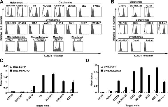
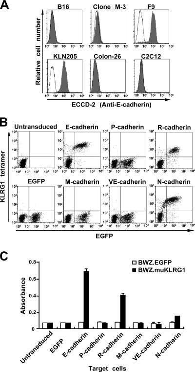
Killer cell lectin-like receptor G1 (KLRG1) is an inhibitory receptor expressed on subsets of natural killer (NK) cells and T cells, for which no endogenous ligands are known. Here, we show that KLRG1 binds three of the classical cadherins (E-, N-, and R-), which are ubiquitously expressed in vertebrates and mediate cell-cell adhesion by homotypic or heterotypic interactions. By expression cloning using the mouse KLRG1 tetramer as a probe, we identified human E-cadherin as a xenogeneic ligand. We also identified a syngeneic interaction between mouse KLRG1 and mouse E-cadherin. Furthermore, we show that KLRG1 binds N- and R-cadherins. Finally, we demonstrate that E-cadherin binding of KLRG1 prevents the lysis of E-cadherin-expressing targets by KLRG1+ NK cells. These results suggest that KLRG1 ligation by E-, N-, or R-cadherins may regulate the cytotoxicity of killer cells to prevent damage to tissues expressing the cadherins.


Figures

Figure 1.
Figure 1.
Expression of a putative KLRG1 ligand on mouse and human cell lines. (A and B) A panel of mouse (A) and human (B) cell lines were stained with mouse KLRG1 tetramer (filled histograms) or PE-streptavidin alone (solid lines) and analyzed by FACS. LLC, Lewis lung cancer. (C and D) The mouse KLRG1 reporter cell line BWZ.muKLRG1(black bars) or the control line BWZ.EGFP (white bars) were cocultured with the indicated mouse (C) or human (D) cell lines and then assayed for β-galactosidase activity. Representative data (means ± SD of triplicate samples) from one of three independent experiments are shown.
Figure 2.
Figure 2.
Expression cloning of a KLRG1 ligand expressed on EBC-1 cells. (A) Biochemical characterization of the putative KLRG1 ligand. The human EBC-1 cell line was surface labeled with 125I, and cell lysates were precipitated with streptavidin-beads loaded with (+) or without (−) biotinylated mouse KLRG1. The precipitates were separated by electrophoresis on an SDS-PAGE gel under reducing (R) or nonreducing (NR) conditions, and 125I-labeled proteins were visualized by phosphorimaging. Arrowheads indicate the bands for the putative KLRG1 ligand. (B) BW5147 cells were transduced with a retroviral cDNA library prepared from EBC-1 cells, and the KLRG1 tetramer+ population was enriched. The enriched KLRG1 tetramer+ population (right) or untransduced control BW5147 cells were stained with the mouse KLRG1 tetramer (filled histograms) or PE-streptavidin alone (solid lines). (C) The KLRG1 tetramer+ population and the control BW5147 cells were tested for stimulation of the KLRG1 reporter cells. Representative data (mean ± SD of triplicate samples) from one of three independent experiments are shown. (D) The KLRG1 tetramer+ population (right) or control BW5147 cells (left) were stained with an anti–human E-cadherin mAb and PE-anti–mouse IgG (filled histograms) or PE-anti–mouse IgG alone (solid lines).
Figure 3.
Figure 3.
Binding of mouse KLRG1 to mouse E-cadherin. (A) BW5147 cells transduced with mouse E-cadherin (right) and control BW5147 cells (left) were analyzed for E-cadherin expression and binding of the KLRG1 tetramer. For E-cadherin expression (top), the cells were stained with mAb to mouse E-cadherin (ECCD-2) and a secondary antibody (filled histograms) or the secondary antibody alone (solid lines). For binding of the mouse KLRG1 tetramer (bottom), the cells were stained with the KLRG1 tetramer (filled histograms) or PE-streptavidin alone (solid lines). (B) BW5147 cells transduced with mouse E-cadherin and control BW5147 cells were tested for stimulation of the KLRG1 reporter cells. Representative data (mean ± SD of triplicate samples) from one of three independent experiments are shown. (C) BW5147 cells transduced with the KLRG1-IRES-EGFP vector (right) or the control EGFP vector (left) were stained with the anti-KLRG1 antibody and PE-anti–mouse IgG. (D) BW5147 cells transduced with the KLRG1-IRES-EGFP vector (bottom) or the control EGFP vector (top) were stained with E-cadherin–Fc fusion protein and PE-anti–human IgG Fc. Where indicated, the cells were treated with the anti-KLRG1 mAb or an isotype-matched control mAb, before staining with the E-cadherin–Fc fusion protein.
Figure 4.
Figure 4.
Binding of KLRG1 to three members of the classical cadherin family. (A) E-cadherin expression on mouse cell lines that bound the KLRG1 tetramer. The indicated cell lines were stained with a mAb to mouse E-cadherin (ECCD-2) and FITC-anti–mouse IgG (filled histograms) or FITC-anti–mouse IgG alone (solid lines). (B and C) BW5147 cells were retrovirally transduced with vectors for bicistronic expression of EGFP and the mouse cadherins indicated. The cells were stained with the KLRG1 tetramer (B) or tested for stimulation of the mouse KLRG1 reporter cells (C). In C, representative data (mean ± SD of triplicate samples) from one of three independent experiments are shown.
Figure 5.
Figure 5.
Ligation of KLRG1 on NK cells by E-cadherin inhibits NK cell cytotoxicity. (A) Expression of KLRG1 on the mouse NK cell line NK03 (left) or the cell line transduced (right) with KLRG1. The cells were stained with the mAb to KLRG1 and a secondary antibody (filled histograms) or the secondary antibody alone (solid lines). (B) Cytotoxicity of the control (left) or KLRG1 transduced (right) mouse NK cell line NK03 against mouse F9 cells was assessed in a 4-h 51Cr-release assay. The assays were conducted in the absence (◯) or the presence (•) of the anti-KLRG1 mAb, or in the presence of an isotype-matched control mAb (▵). (C) Cytotoxicity of the control (left) or KLRG1 transduced (right) mouse NK cells against E-cadherin expressing (bottom) or control (top) BW5147 targets, both of which lack Fcγ receptors. The assays were conducted in the absence (◯) or the presence (•) of the anti-KLRG1 mAb, or in the presence of an isotype-matched control mAb (▵). Representative data (mean ± SD of triplicate samples) from one of three independent experiments are shown.

Comment in

References

    1. Biron, C.A., K.B. Nguyen, G.C. Pien, L.P. Cousens, and T.P. Salazar-Mather. 1999. Natural killer cells in antiviral defense: function and regulation by innate cytokines. Annu. Rev. Immunol. 17:189–220. - PubMed
    1. Lanier, L.L. 2005. NK cell recognition. Annu. Rev. Immunol. 23:225–274. - PubMed
    1. Long, E.O. 1999. Regulation of immune responses through inhibitory receptors. Annu. Rev. Immunol. 17:875–904. - PubMed
    1. Moretta, L., and A. Moretta. 2004. Killer immunoglobulin-like receptors. Curr. Opin. Immunol. 16:626–633. - PubMed
    1. Yokoyama, W.M. 1998. Natural killer cell receptors. Curr. Opin. Immunol. 10:298–305. - PubMed

Publication types

MeSH terms