. 2006 Feb 20;203(2):383-91.
doi: 10.1084/jem.20052256. Epub 2006 Feb 6.

H2-M3-restricted CD8+ T cells are not required for MHC class Ib-restricted immunity against Listeria monocytogenes

Affiliations

H2-M3-restricted CD8+ T cells are not required for MHC class Ib-restricted immunity against Listeria monocytogenes

Sarah E F D'Orazio et al. J Exp Med. .

Abstract

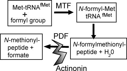
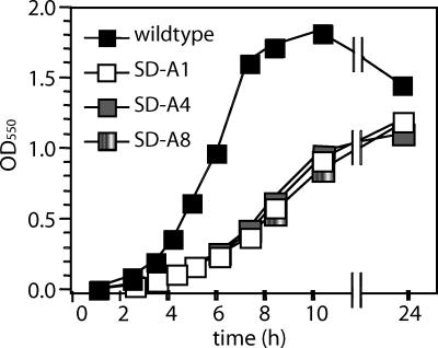
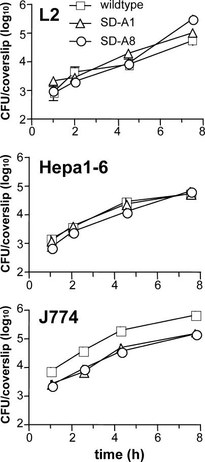
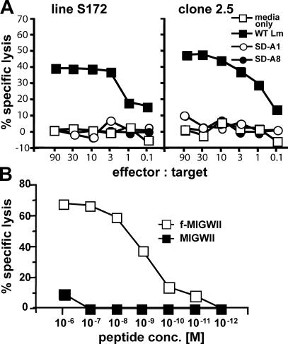
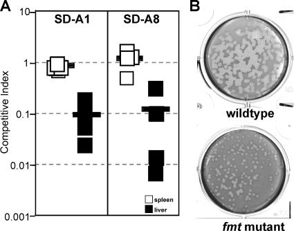
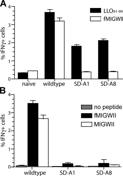
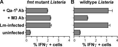
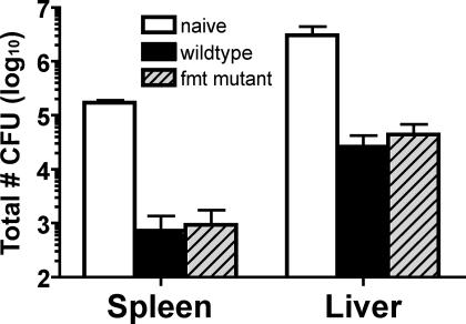
Studies using major histocompatibility complex (MHC)-Ia-deficient mice have shown that MHC-Ib-restricted CD8+ T cells can clear infections caused by intracellular pathogens such as Listeria monocytogenes. M3-restricted CD8+ T cells, which recognize short hydrophobic N-formylated peptides, appear to comprise a substantial portion of the MHC-Ib-restricted T cell response in the mouse model of L. monocytogenes infection. In this study, we isolated formyltransferase (fmt) mutant strains of L. monocytogenes that lacked the ability to add formyl groups to nascent polypeptides. These fmt mutant Listeria strains did not produce antigens that could be recognized by M3-restricted T cells. We showed that immunization of MHC-Ia-deficient mice with fmt mutant Listeria resulted in stimulation of a protective memory response that cleared subsequent challenge with wild-type L. monocytogenes, despite the fact that M3-restricted CD8+ T cells did not proliferate in these mice. These data suggest that M3-restricted T cells are not required for protection against L. monocytogenes and underscore the importance of searching for new antigen-presenting molecules among the large MHC-Ib family of proteins.


Figures

Figure 1.
Figure 1.
Actinonin inhibits bacterial peptide deformylase (PDF) activity. Bacterial methionyl-tRNAfMet formyltransferase (MTF) adds a formyl group to methionines that are bound to initiator tRNAs. Formylation promotes binding of initiator factor 2 (IF-2) and impairs binding of elongation factor Tu and, therefore, provides a basis for the initiation of protein synthesis. The formyl group is cleaved from the mature protein by peptide deformylase (PDF), and in many cases the NH2-terminal methionine is removed by methionine amino peptidase (not depicted). Actinonin is a naturally occurring inhibitor of metallohydrolases that blocks PDF activity in bacteria (reference 20).
Figure 2.
Figure 2.
The doubling time of fmt mutant Listeria is increased compared with wild-type Listeria during growth in BHI broth. Freshly streaked wild-type (Lm 10403s) or fmt mutant (SD-A1, SD-A4, SD-A8) L. monocytogenes strains were incubated overnight at 37°C with aeration in BHI broth. The next day, each culture was diluted to an OD550 of ∼0.015 in fresh BHI broth and further incubated at 37°C with aeration. Bacterial growth was assessed by determining the OD550 at various time points as indicated. Representative data from one of two different experiments are shown.
Figure 3.
Figure 3.
fmt mutant Listeria do not have a replication defect during intracellular growth. Monolayers of L2 fibroblasts, Hepa1-6 epithelial cells, or J774 macrophage-like cells plated on glass coverslips were infected with the Listeria strains indicated for 30 min, washed ex-tensively, and incubated in media containing 25 μg/ml gentamicin to kill extracellular bacteria. At each of the time points indicated, the cells at-tached to each coverslip were lysed in sterile water to release intracellular bacteria. Serial dilutions of each lysate were plated on BHI agar to deter-mine the total number of bacterial colony forming units (CFU) associated with each coverslip. Data shown represent the average values ± SD for triplicate coverslips from one of two separate experiments.
Figure 4.
Figure 4.
Formylated peptides do not accumulate in the culture supernatants of fmt mutant Listeria strains. T cell recognition of target cells was measured as a function of cytotoxicity (51Cr release into the supernatant) and is shown as percentage of specific lysis. (A) 51Cr-loaded EL-4 target cells were pretreated with media alone (BHI broth) or culture supernatant derived from either wild-type (wt Lm) or fmt mutant (SD-A1, SD-A8) L. monocytogenes strains. CD8+ T cells were added at various effector to target ratios and the cells were coincubated for 4 h. Line S172 T cells recognize N-formylated MIGWII peptide bound to M3; clone 2.5 is an M3-restricted CD8+ T clone of unknown specificity. (B) 51Cr-loaded EL-4 target cells were pretreated with synthetic peptide at the final concentrations indicated. Line S172 T cells were added at an effector: target ratio of 45:1.
Figure 5.
Figure 5.
fmt mutant Listeria are less virulent in the liver than in the spleen. (A) A competitive index assay was used to determine the relative virulence of fmt mutant Listeria strains SD-A1 and SD-A8 compared with wild-type Listeria. BALB/c mice were sublethally infected intravenously with a 1:1 mixture of wild-type (erythromycin-resistant) and mutant Listeria (erythromycin-sensitive). Mice were killed 3 d later and the spleens and livers were homogenized and differentially plated to determine the total number of each bacterial strain present. Squares represent competitive indices for individual mice; thick black lines represent average indices for groups of mice. (B) A plaque assay was used to assess the degree of cell-to-spread achieved by fmt mutant Lm SD-A1 compared with wild-type Lm 10403s. Confluent monolayers of L2 fibroblasts were infected at low MOI (0.1) for 1 h and agarose overlays were added. After 3 d of incubation at 37°C in 7% CO2, neutral red overlays were added to visualize the plaques. Plaque size indicates the relative degree of cell-to-cell spread for each strain. Infections were performed in duplicate wells in two separate experiments.
Figure 6.
Figure 6.
M3-restricted CD8+ T cells are not activated during infection with fmt mutant Listeria. Mice were immunized with PBS (naive group) or a sublethal dose of either wild-type (10403s) or fmt mutant (SD-A1, SD-A8) L. monocytogenes. 7 d later, the mice were killed and the number of antigen-specific CD8+ T cells present in the spleen was determined by IFNγ ICCS. (A) BALB/c splenocytes were exposed to either 1 μM LLO91-99 peptide, which binds to Kd (black bars) or 1 μM fMIGWII peptide, which binds to M3 (white bars). (B) MHC-Ia–deficient splenocytes were incubated with either no peptide (gray bars), 1 μM formylated fMIGWII peptide (black bars), or 1 μM unformylated MIGWII peptide (white bars). Data shown represent the average number of CD8β + cells that were IFNγ+ within each group of mice ± SE from one of three separate experiments.
Figure 7.
Figure 7.
CD8+ T cells from fmt mutant Listeria-immune mice recognize antigens presented by an MHC-Ib molecule other than M3 or Qa-1b. Groups of MHC-Ia–deficient mice were infected with 2,000 CFU of either (A) fmt mutant (Lm SD-A1) or (B) wild-type (Lm 10403s) L. monocytogenes. 7 d after infection, splenocytes from these mice were harvested and exposed to MHC-Ia–deficient BMMΦ (either uninfected or cells that had been infected for 3 h with the same strain of Listeria used to infect the mice). As indicated, some samples of Listeria-infected BMMΦ were pretreated with either anti-M3 or anti–Qa-1b monoclonal antibody before the addition of immune splenocytes. Data shown represent the average number of TCRβ+ CD8+ cells that stained IFNγ+ within each group of mice ± SE from one out of two experiments.
Figure 8.
Figure 8.
Immunization with fmt mutant Listeria results in full protective immunity against secondary challenge with wild-type Listeria. Groups of MHC-Ia–deficient mice were immunized with PBS (naive) or 103 CFU of either 10403s (wild type) or SD-A1 (fmt mutant) L. monocytogenes. 3 wk later, all groups of mice were challenged intravenously with 5 × 10⁴ wild-type Listeria. The total number of CFU per organ was determined 3 d after infection. Similar results were observed for groups of mice immunized with strain SD-A8 (not depicted). Data shown represent average values ± SD for groups of mice from one of two independent experiments.

Similar articles

Cited by

References

    1. Busch, D.H., K. Kerksiek, and E.G. Pamer. 1999. Processing of Listeria monocytogenes antigens and the in vivo T-cell response to bacterial infection. Immunol. Rev. 172:163–169. - PubMed
    1. Geginat, G., S. Schenk, M. Skoberne, W. Goebel, and H. Hof. 2001. A novel approach of direct ex vivo epitope mapping identifies dominant and subdominant CD4 and CD8 T cell epitopes from Listeria monocytogenes. J. Immunol. 166:1877–1884. - PubMed
    1. Soloski, M.J., M.E. Szperka, A. Davies, and S.L. Wooden. 2000. Host immune response to intracellular bacteria: a role for MHC-linked class-Ib antigen-presenting molecules. Proc. Soc. Exp. Biol. Med. 224:231–239. - PubMed
    1. Forman, J., and K.F. Lindahl. 2002. Listing, Location, Binding Motifs, and Expression of Nonclassical Class I and Related Genes and Molecules. In Current Protocols in Immunology. John Wiley & Sons, Hoboken, NJ. A.1M1–A.1M13. - PubMed
    1. Shawar, S.M., J.M. Vyas, J.R. Rodgers, and R.R. Rich. 1994. Antigen presentation by major histocompatibility complex class Ib molecules. Annu. Rev. Immunol. 12:839–880. - PubMed

Publication types

MeSH terms