. 2006 Feb 13;172(4):505-15.
doi: 10.1083/jcb.200510028. Epub 2006 Feb 6.

NEDD1-dependent recruitment of the gamma-tubulin ring complex to the centrosome is necessary for centriole duplication and spindle assembly

Affiliations

NEDD1-dependent recruitment of the gamma-tubulin ring complex to the centrosome is necessary for centriole duplication and spindle assembly

Laurence Haren et al. J Cell Biol. .

Abstract

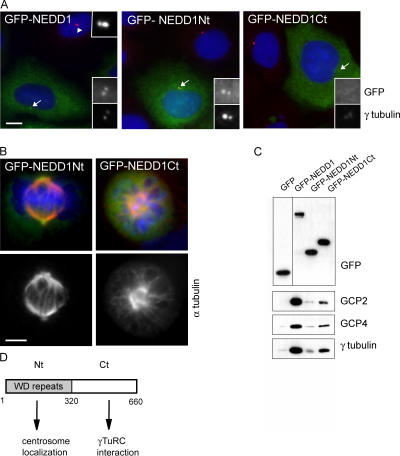
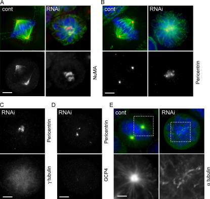
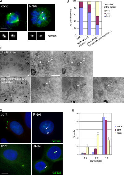
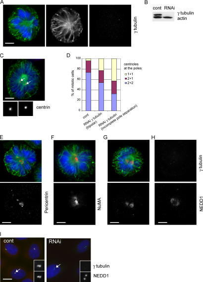
The centrosome is the major microtubule organizing structure in somatic cells. Centrosomal microtubule nucleation depends on the protein gamma-tubulin. In mammals, gamma-tubulin associates with additional proteins into a large complex, the gamma-tubulin ring complex (gammaTuRC). We characterize NEDD1, a centrosomal protein that associates with gammaTuRCs. We show that the majority of gammaTuRCs assemble even after NEDD1 depletion but require NEDD1 for centrosomal targeting. In contrast, NEDD1 can target to the centrosome in the absence of gamma-tubulin. NEDD1-depleted cells show defects in centrosomal microtubule nucleation and form aberrant mitotic spindles with poorly separated poles. Similar spindle defects are obtained by overexpression of a fusion protein of GFP tagged to the carboxy-terminal half of NEDD1, which mediates binding to gammaTuRCs. Further, we show that depletion of NEDD1 inhibits centriole duplication, as does depletion of gamma-tubulin. Our data suggest that centriole duplication requires NEDD1-dependent recruitment of gamma-tubulin to the centrosome.


Figures

Figure 1.
Figure 1.
NEDD1 is recruited to the mitotic centrosome and to spindle microtubules and colocalizes with γ-tubulin. (A) Alignment of the WD repeats of human NEDD1 (available from GenBank/EMBL/DDBJ under accession no. 74762597) and Dgp71WD (accession no. 28628541), together with those of human transducin β chain 1 (GBB; accession no. 51317302). The positions of predicted β strands (A, B, C, and D) are indicated above the alignment. Green and orange boxes indicate conservation of hydrophobic and aromatic residues, respectively. Additional sequence hallmarks found in WD repeats are indicated in yellow, red, and blue. (B–E) HeLa cells immunostained for NEDD1 (red), α-tubulin (green), and DNA (blue). NEDD1 is enriched at the mitotic centrosome and is found along spindle and midbody microtubules (arrows). (B) Interphase and prometaphase cells; (C) metaphase cell; (D) anaphase cells; (E) telophase cell. Bar, 5 μm. (F–H) Cells stained for NEDD1 (red), γ-tubulin (green), and DNA (blue). NEDD1 colocalizes with γ-tubulin throughout cell cycle, at interphase (F), metaphase (G), and telophase (H). Bar,5 μm. (I) HeLa cell lysates immunoblotted for NEDD1: untreated cells (cont), after overnight incubation in 500 nM taxol (mitotic arrest), or lysate of cells in mitotic arrest treated with λ-phosphatase (400 U for 30 min at 30°C).
Figure 2.
Figure 2.
Silencing of NEDD1 induces mitotic defects. HeLa cells were treated without siRNA (mock), with control siRNA (cont), or with NEDD1 siRNA (RNAi) for various time points. (A) Immunoblot of crude cell lysates (40 μg) showing reduction of NEDD1 levels but no γ-tubulin reduction. NEDD1 levels in mock-treated cells diluted 1/5 and 1/10 are presented to compare with depleted cells. (B) Histogram indicating percentage of mitotic cells at different stages of mitosis (mean of three experiments ± SEM; 350–400 total cells scored per condition). Most of the cells are arrested in a prometaphase-like state with poorly separated spindle poles. (C–G) Mitotic cells stained for NEDD1 (red), α-tubulin (green), and DNA (blue). Bar, 5 μm.
Figure 3.
Figure 3.
Silencing of NEDD1 inhibits recruitment of the γTuRC to the mitotic centrosome, but the majority of γTuRCs can still form. (A) NEDD1-depleted HeLa cells (RNAi) or cells treated with control siRNA (cont) that were stained for NEDD1 (green), γ-tubulin (red), and DNA (blue). (B–D) Cells stained for α-tubulin (green) and γ-tubulin, GCP2 or -4 (red), and DNA (blue). Bars, 5 μm. (E) Coimmunoprecipitation of NEDD1 and γTuRC proteins. (left) Immunoprecipitation from HeLa cells using antibodies against NuMA or NEDD1. (right) Immunoprecipitation with polyclonal antibody R75 against γ-tubulin or with preimmune serum from the same animal (preim.). Precipitates w ere separated by gel electrophoresis, blotted, and probed with antibodies against GCP2, GCP4, NEDD1, and γ-tubulin. (F) Coimmunoprecipitation of γTuRC proteins from cells treated with control siRNA or with siRNA against NEDD1 (RNAi). Conditions were as in the right column of E. (G) Comparison of extracts from HeLa cells treated with control and NEDD1 siRNA (RNAi) after fractionation in gradients of 5–40% sucrose. The fractions were precipitated using methanol and immunoblotted with antibodies against GCP2, GCP4, NEDD1, and γ-tubulin. Arrowheads indicate the position of the 32S γTuRC, and 19S indicates the sedimentation of thyroglobulin. (H) Coimmunoprecipitation of NEDD1 and γ-tubulin from the purified 32S fraction of undepleted extracts, using the polyclonal antibody R75 against γ-tubulin and control precipitation with preimmune serum from the same animal.
Figure 4.
Figure 4.
Silencing of NEDD1 in interphase cells affects centrosomal localization of γ-tubulin and microtubule nucleation. (A and B) HeLa cells treated with control siRNA (cont) or NEDD1 siRNA (RNAi) and stained for NEDD1 or pericentrin (green), γ-tubulin (red), and DNA (blue). Centrosomes appear in yellow in control cells because NEDD1 or pericentrin colocalize with γ-tubulin. Insets show enlarged views of the centrosomes indicated by arrows. (C) Microtubule regrowth assay in U2OS cells treated with control siRNA or in cells treated with siRNA against NEDD1. Cells were cold treated and reheated for 0, 1, or 2 min at 37°C before staining for NEDD1 (green), α-tubulin (red), and DNA (blue). Arrows point to microtubule asters nucleated in control cells; in RNAi-treated cells, asters are barely visible. Bars, 10 μm.
Figure 5.
Figure 5.
The carboxy-terminal domain of NEDD1 binds to γTuRCs, and the amino-terminal domain mediates centrosome attachment. (A) Transient overexpression of GFP fusion proteins with full-length NEDD1 (GFP-NEDD1) and amino acids 1–319 (GFP-NEDD1Nt) and 321–660 (GFP-NEDD1Ct) in U2OS cells. Cells are stained for γ-tubulin (red) and DNA (blue). Arrows indicate the positions of centrosomes that are shown enlarged in insets, displaying the fluorescence of the various GFP fusion proteins or γ-tubulin. For comparison, an untransfected cell is indicated by an arrowhead in the left panel (inset in top right corner shows an enlarged view). Bar, 10 μm. (B) Mitotic U2OS cells overexpressing GFP-NEDD1Nt or GFP-NEDD1Ct (green) that were stained for α-tubulin (red) and DNA (blue). GFP-NEDD1Ct induces spindle defects. Bar, 5 μm. (C) Immunoprecipitation of the overexpressed proteins by anti-GFP antibody in Cos-7 cells. (top) Detection of precipitated proteins by anti-GFP antibody (note that GFP is resolved in a 10% polyacrylamide gel, whereas the other proteins are separated in a 7.5% gel). (bottom) γTuRC proteins (γ-tubulin, GCP2, and GCP4) coprecipitate with GFP-NEDD1 and GFP-NEDD1Ct. (D) Schematic drawing depicting functional domains of NEDD1.
Figure 6.
Figure 6.
Dispersion of the microtubule organization centers in NEDD1-depleted mitotic cells. (A and B) HeLa cells stained for α-tubulin (green) and DNA (blue), NuMA (A, red) as a marker of microtubule minus ends, and pericentrin (B, red) as a marker of the pericentriolar material. NEDD1-depleted cells (RNAi) show poorly separated and disorganized spindle poles. (C and D) NEDD1-depleted cells stained for pericentrin and the γTuRC proteins γ-tubulin (C) and GCP4 (D). (E) Microtubule regrowth assay in mitotic cells. Cells were cold treated and reheated for 30 s at 37°C before staining for NEDD1 (green), α-tubulin (red), and DNA (blue). Magnified areas show microtubules nucleated from the centrosome in control cells and from dispersed sites in silenced cells. Bars, 5 μm.
Figure 7.
Figure 7.
Silencing of NEDD1 inhibits centriole duplication. (A) HeLa cells stained for α-tubulin (green), DNA (blue), and centrin as a marker of the centrioles (red and insets). Cells that were efficiently depleted of NEDD1 (RNAi) show poorly separated poles, containing only one centriole at each pole. Arrows indicate positions of the centrosomes. Bar, 5 μm. (B) Quantification of the number of centrin-stained centrioles per pole (mean of three experiments; 228 control RNA-treated cells, 138 NEDD1-depleted cells with fully separated poles, and 322 depleted cells with incompletely separated poles were scored). (C) Electron microscopy of a cell with a bipolar mitotic spindle (RNAi bipolar) and a depleted cell with a monopolar phenotype (RNAi incomplete pole separation). The pictures on the left show low magnifications, and selected areas at high magnifications are at the right. Serial sections of spindle poles reveal a pair of closely associated centrioles (1 and 2) oriented at a 90° angle in the bipolar spindle, whereas only a single centriole (1) could be found at the pole of the depleted cell. Bar, 500 nm. (D) U2OS cells treated with aphidicolin and stained for NEDD1 (red), DNA (blue), and centrin or polyglutamylated tubulin (GT335; green). Control cells or poorly depleted cells (arrowhead in bottom right image) show clouds of numerous centriole dots, whereas efficiently depleted cells have mainly 1–4 centrioles (arrows). Bar, 10 μm. (E) Quantification of centriole numbers per cell was based on centrin staining. Numbers were obtained from selected cells that displayed a strong reduction of NEDD1 levels (mean of three experiments ± SEM; 350–400 cells scored per condition).
Figure 8.
Figure 8.
Silencing of γ-tubulin inhibits centriole duplication and causes spindle defects similar to those of NEDD1 depletion. (A) Mitotic HeLa cell depleted of γ-tubulin and stained for α-tubulin (green), γ-tubulin (red), and DNA (blue). Bar, 5 μm. (B) Immunoblot of lysates (40 μg) of cells treated with control siRNA (cont) or γ-tubulin siRNA (RNAi) and probed with antibodies against γ-tubulin and actin. (C) A depleted cell stained for centrin (red), α-tubulin (green), and DNA (blue). Insets show magnified views of centrin staining of left and right spindle poles, respectively. Arrows indicate positions of the centrosomes. Bar, 5 μm. (D) Quantification of centrin-stained centrioles per pole (mean of two experiments; 232 control RNA-treated cells, 35 γ-tubulin–depleted cells with fully separated poles, and 180 depleted cells with incompletely separated poles were scored). (E–G) Cells depleted of γ-tubulin and stained in green for α-tubulin and in red for pericentrin (E), NuMA (F), and NEDD1 (G). (H) Cell depleted of γ-tubulin and stained for γ-tubulin and NEDD1. Mitotic cells depleted of γ-tubulin show disorganized spindle poles and diffuse staining of NEDD1. (I) Control and γ-tubulin–depleted interphase cells (RNAi) stained for γ-tubulin (red), NEDD1 (green), and DNA (blue). NEDD1 is concentrated at the centrosome (arrows and bottom insets), even in the absence of γ-tubulin (top insets).

References

    1. Andersen, J.S., C.J. Wilkinson, T. Mayor, P. Mortensen, E.A. Nigg, and M. Mann. 2003. Proteomic characterization of the human centrosome by protein correlation profiling. Nature. 426:570–574. - PubMed
    1. Balczon, R., L. Bao, W.E. Zimmer, K. Brown, R.P. Zinkowski, and B.R. Brinkley. 1995. Dissociation of centrosome replication events from cycles of DNA synthesis and mitotic division in hydroxyurea-arrested Chinese hamster ovary cells. J. Cell Biol. 130:105–115. - PMC - PubMed
    1. Barbosa, V., R.R. Yamamoto, D.S. Henderson, and D.M. Glover. 2000. Mutation of a Drosophila gamma tubulin ring complex subunit encoded by discs degenerate-4 differentially disrupts centrosomal protein localization. Genes Dev. 14:3126–3139. - PMC - PubMed
    1. Barbosa, V., M. Gatt, E. Rebollo, C. Gonzalez, and D.M. Glover. 2003. Drosophila dd4 mutants reveal that gammaTuRC is required to maintain juxtaposed half spindles in spermatocytes. J. Cell Sci. 116:929–941. - PubMed
    1. Callebaut, I., G. Labesse, P. Durand, A. Poupon, L. Canard, J. Chomilier, B. Henrissat, and J.P. Mornon. 1997. Deciphering protein sequence information through hydrophobic cluster analysis (HCA): current status and perspectives. Cell. Mol. Life Sci. 53:621–645. - PMC - PubMed

Publication types