. 2006 Aug;291(2):L166-74.
doi: 10.1152/ajplung.00160.2005. Epub 2006 Feb 3.

Cyclic stretch attenuates effects of hyperoxia on cell proliferation and viability in human alveolar epithelial cells

Affiliations

Cyclic stretch attenuates effects of hyperoxia on cell proliferation and viability in human alveolar epithelial cells

Ryan M McAdams et al. Am J Physiol Lung Cell Mol Physiol. 2006 Aug.

Abstract

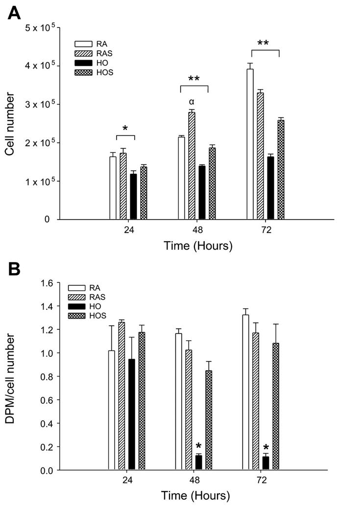
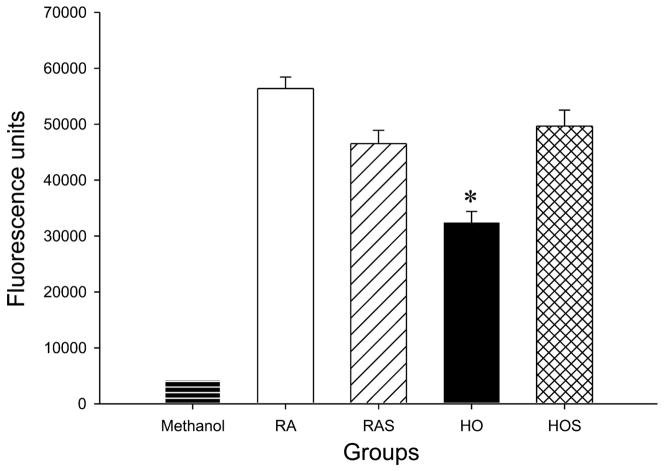
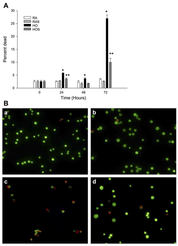
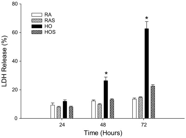
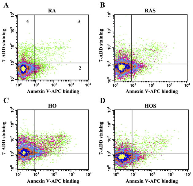
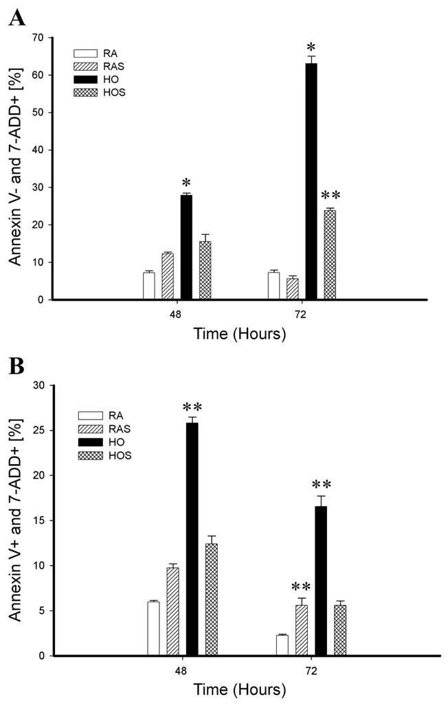
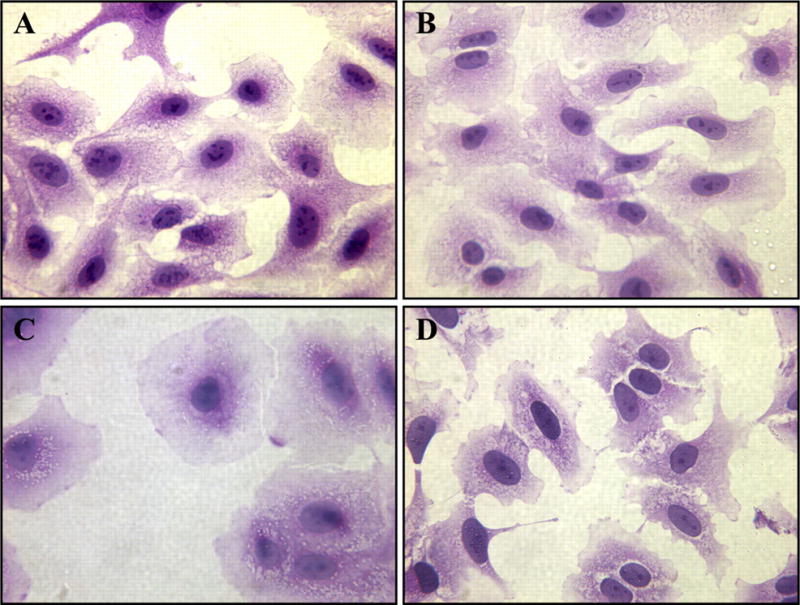
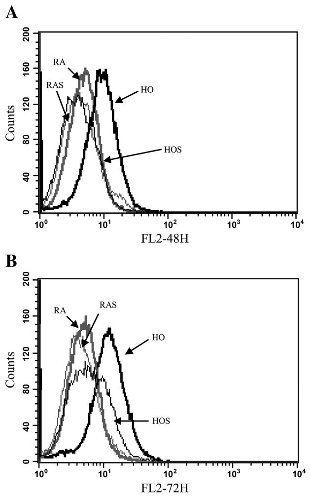
The treatment of severe lung disease often requires the use of high concentrations of oxygen coupled with the need for assisted ventilation, potentially exposing the pulmonary epithelium to both reactive oxygen species and nonphysiological cyclic stretch. Whereas prolonged hyperoxia is known to cause increased cell injury, cyclic stretch may result in either cell proliferation or injury depending on the pattern and degree of exposure to mechanical deformation. How hyperoxia and cyclic stretch interact to affect the pulmonary epithelium in vitro has not been previously investigated. This study was performed using human alveolar epithelial A549 cells to explore the combined effects of cyclic stretch and hyperoxia on cell proliferation and viability. Under room air conditions, cyclic stretch did not alter cell viability at any time point and increased cell number after 48 h compared with unstretched controls. After exposure to prolonged hyperoxia, cell number and [(3)H]thymidine incorporation markedly decreased, whereas evidence of oxidative stress and nonapoptotic cell death increased. The combination of cyclic stretch with hyperoxia significantly mitigated the negative effects of prolonged hyperoxia alone on measures of cell proliferation and viability. In addition, cyclic stretch resulted in decreased levels of oxidative stress over time in hyperoxia-exposed cells. Our results suggest that cyclic stretch, as applied in this study, can minimize the detrimental effects of hyperoxia on alveolar epithelial A549 cells.


Figures

Fig. 1
Fig. 1
A: effects of hyperoxia and stretch on cell number. A549 cells were exposed to room air (RA), room air with stretch (RAS), hyperoxia (HO), and hyperoxia with stretch (HOS) for 24, 48, and 72 h. Cell number was determined by manually counting cells from duplicate chambers with a hemacytometer. Values are expressed as means ± SE, with n = 3 independent experiments. *P < 0.05, RA and RAS vs. HO. **P < 0.05, RA, RAS, and HOS vs. HO. αP < 0.05, RA vs. RAS. B: effects of hyperoxia and stretch on [3H]thymidine incorporation. A549 cells were exposed to RA, RAS, HO, and HOS for 24, 48, and 72 h. Cell proliferation was assessed as [3H]thymidine incorporation corrected for cell number. Cells were pulsed for 12 h with 1 μCi/ml [3H]thymidine and analyzed using a liquid scintillation analyzer. Values are expressed as means ± SE, with n = 3 independent experiments. *P < 0.05, RA, RAS, and HOS vs. HO. DPM, disintegrations per minute.
Fig. 2
Fig. 2
Effects of hyperoxia and stretch on cell viability. A549 cells were exposed to RA, RAS, HO, and HOS for 24, 48, and 72 h. Cell suspensions were transferred to 96-well flat-bottomed microtiter plates, and 25 μM calcein AM was added to each well. Cells were incubated in the dark and analyzed using a spectrophotometer plate reader at an excitation wavelength of ~485 nm and an emission wavelength of ~530 nm. Methanol-exposed cells serve as positive controls for maximal cell death (lowest viability). Values are expressed as means ± SE, with n = 3 independent experiments. *P < 0.05, HO vs. RA, RAS, and HOS.
Fig. 3
Fig. 3
A: effects of hyperoxia and stretch on cell viability and death. A549 cells were exposed to RA, RAS, HO, and HOS for 24, 48, and 72 h. With the use of a LIVE/DEAD assay, cell suspensions were transferred to 96-well microtiter assay plates. Positive control wells were treated with 100 μl of methanol to induce maximal death. Ethidium homodimer-1 (EthD-1; 17 μM) and calcein AM (10 μM) were added to the wells, and the cells were incubated in the dark and then analyzed using fluorescence microscopy. Cell counts were made on the basis of the number of live cells (green) vs. dead cells (red) that were seen in random, noncontiguous fields until ~250 cells total were counted. Values are expressed as means ± SE, with n = 3 independent experiments. *P < 0.05, HO vs. RA, RAS, and HOS. **P < 0.05, HOS vs. RA and RAS. B: effects of hyperoxia and stretch on cell viability and death. Fluorescent microscopy images represent the following groups of A549 cells after 72 h of exposure to RA (a), RAS (b), HO (c), and HOS (d). Cells were stained with calcein AM, which produces a bright green fluorescence in viable cells, and EthD-1, which produces a red fluorescence reflective of cell death. Scale bar, 100 μm. Images represent typical findings from 3 independent studies.
Fig. 4
Fig. 4
Lactate dehydrogenase (LDH) release assay. A549 cells were exposed to RA, RAS, HO, and HOS for 24, 48, and 72 h. Samples were assayed for LDH release, a marker of cell death, in culture medium. Values are expressed as means ± SE, with n = 3 independent experiments. *P < 0.01, HO vs. RA, RAS, and HOS.
Fig. 5
Fig. 5
Determination of apoptosis and necrosis. Cells were stretched, harvested at 72 h, stained with annexin V-APC and 7-aminoactinomycin (7-ADD) and analyzed using flow cytometry. Plots of flow cytometry analysis represent the following groups: RA (A), RAS (B), HO (C), and HOS (D). Intensity of 7-ADD staining (y-axis) is plotted vs. annexin V-APC intensity (x-axis). In all 4 plots, viable cells appear at bottom left (quadrant 1, annexin V negative/7-ADD negative), early apoptotic cells at bottom right (quadrant 2, annexin V positive/7-ADD negative), late apoptotic/necrotic cells at top right (quadrant 3, annexin V positive/7-ADD positive), and necrotic cells at top left (quadrant 4, annexin V negative/7-ADD positive).
Fig. 6
Fig. 6
Annexin V-APC binding and 7-ADD staining. Flow cytometry data of A549 cells exposed to RA, RAS, HO, and HOS represent a comparison between groups at 48 and 72 h of exposure. A: the percentage of necrotic cells (annexin V-APC − and 7-ADD +). *P < 0.01, HO > RA, RAS, and HOS. **P < 0.05 HOS > RA and RAS. B: the percentage of late apoptotic/necrotic cells (annexin V-APC + and 7-ADD −). **P < 0.05, RAS > RA and HO > RA, RAS, and HOS.
Fig. 7
Fig. 7
Effects of hyperoxia and stretch on A549 cell morphology. After 72 h of exposure to RA (A), RAS (B), HO (C), and HOS (D), A549 cells were stained with hematoxylin and eosin as described in METHODS. Scale bar, 50 μm.
Fig. 8
Fig. 8
Superoxide detection with dihydroethidium (DHE) staining using flow cytometry. A549 cells were stained with DHE after 48 (A) and 72 h (B) of exposure to RA (thick shaded line), RAS (thin shaded line), HO (thick solid line), and HOS (thin solid line) to detect superoxide. Histogram overlay represents comparisons between groups after 24, 48, and 72 h of exposure. A right shift in the FL2 channel detecting orange-red fluorescence represents the presence of superoxide (nuclear binding of ethidium). P < 0.05, HO vs. RA, RAS, and HOS. P < 0.05, HOS vs. RA and RAS.

Similar articles

Cited by

References

    1. Allen RG, Tresini M. Oxidative stress and gene regulation. Free Radic Biol Med. 2000;28:463–499. - PubMed
    1. Cavanaugh KJ, Jr, Margulies SS. Measurement of stretch-induced loss of alveolar epithelial barrier integrity with a novel in vitro method. Am J Physiol Cell Physiol. 2002;283:C1801–C1808. - PubMed
    1. Chang L, Karin M. Mammalian MAP kinase signalling cascades. Nature. 2001;410:37–40. - PubMed
    1. Chess PR, Toia L, Finkelstein JN. Mechanical strain-induced proliferation and signaling in pulmonary epithelial H441 cells. Am J Physiol Lung Cell Mol Physiol. 2000;279:L43–L51. - PubMed
    1. Dos Santos CC, Slutsky AS. Mechanisms of ventilator-induced lung injury: a perspective. J Appl Physiol. 2000;89:1645–1655. - PubMed

LinkOut - more resources