

Unique, common, and interacting cortical correlates of thirst and pain
- PMID: 16461454
- PMCID: PMC1413749
- DOI: 10.1073/pnas.0511019103
Unique, common, and interacting cortical correlates of thirst and pain
Abstract
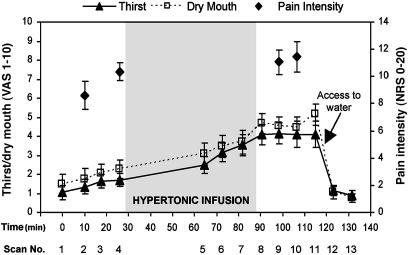
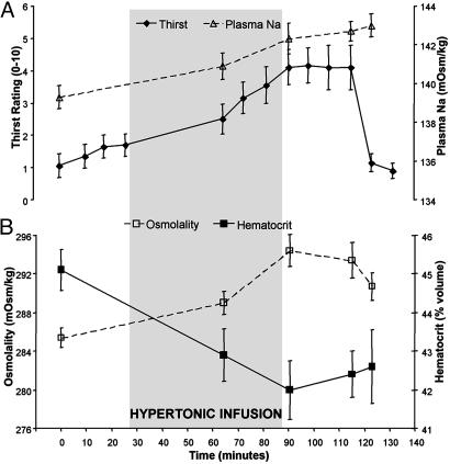
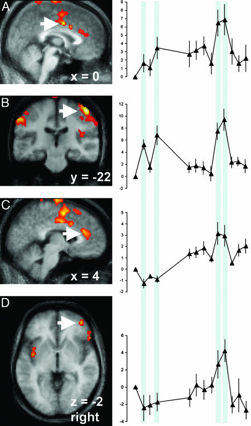
This study used positron-emission tomography to establish the patterns of brain activity involved in the isolated and concurrent experiences of thirst and pain. Ten subjects were scanned while experiencing pain evoked with noxious pressure, while experiencing thirst after the infusion of hypertonic saline, and while experiencing pain when thirsty. After the onset of thirst, noxious pressure evoked more intense sensations of pain. Noxious pressure did not change subjective ratings of thirst. Thirst caused activation in the anterior cingulate (Brodmann area 32) and the insula. Enhanced pain responses were associated with increased activity in cortical regions that are known to correlate with pain intensity, and also with unique activity in the pregenual anterior cingulate and ventral orbitofrontal cortex. These findings suggest a role for limbic and prefrontal cortices in the modulation of pain during the experience of thirst.
Conflict of interest statement
Conflict of interest statement: No conflicts declared.
Figures

Similar articles
-
Paralimbic and medial prefrontal cortical involvement in neuroendocrine responses to traumatic stimuli.Am J Psychiatry. 2007 Aug;164(8):1250-8. doi: 10.1176/appi.ajp.2007.06081367. Am J Psychiatry. 2007. PMID: 17671289
-
Modulation of cortical-limbic pathways in major depression: treatment-specific effects of cognitive behavior therapy.Arch Gen Psychiatry. 2004 Jan;61(1):34-41. doi: 10.1001/archpsyc.61.1.34. Arch Gen Psychiatry. 2004. PMID: 14706942
-
Human cortical responses to water in the mouth, and the effects of thirst.J Neurophysiol. 2003 Sep;90(3):1865-76. doi: 10.1152/jn.00297.2003. Epub 2003 May 28. J Neurophysiol. 2003. PMID: 12773496
-
The contribution of neuroimaging techniques to the understanding of supraspinal pain circuits: implications for orofacial pain.Oral Surg Oral Med Oral Pathol Oral Radiol Endod. 2005 Sep;100(3):308-14. doi: 10.1016/j.tripleo.2004.11.014. Oral Surg Oral Med Oral Pathol Oral Radiol Endod. 2005. PMID: 16122658 Review.
-
Cortico-limbic pain mechanisms.Neurosci Lett. 2019 May 29;702:15-23. doi: 10.1016/j.neulet.2018.11.037. Epub 2018 Nov 29. Neurosci Lett. 2019. PMID: 30503916 Free PMC article. Review.
Cited by
-
Corticospinal and peripheral responses to heat-induced hypo-hydration: potential physiological mechanisms and implications for neuromuscular function.Eur J Appl Physiol. 2022 Aug;122(8):1797-1810. doi: 10.1007/s00421-022-04937-z. Epub 2022 Apr 1. Eur J Appl Physiol. 2022. PMID: 35362800 Free PMC article. Review.
-
Regional brain responses associated with using imagination to evoke and satiate thirst.Proc Natl Acad Sci U S A. 2020 Jun 16;117(24):13750-13756. doi: 10.1073/pnas.2002825117. Epub 2020 Jun 1. Proc Natl Acad Sci U S A. 2020. PMID: 32482871 Free PMC article.
-
Do Individual Differences in Perception Affect Awareness of Climate Change?Brain Sci. 2024 Mar 9;14(3):266. doi: 10.3390/brainsci14030266. Brain Sci. 2024. PMID: 38539654 Free PMC article.
-
Psychosocial versus physiological stress - Meta-analyses on deactivations and activations of the neural correlates of stress reactions.Neuroimage. 2015 Oct 1;119:235-51. doi: 10.1016/j.neuroimage.2015.06.059. Epub 2015 Jun 26. Neuroimage. 2015. PMID: 26123376 Free PMC article.
-
Thirst and the state-dependent representation of incentive stimulus value in human motive circuitry.Soc Cogn Affect Neurosci. 2015 Dec;10(12):1722-9. doi: 10.1093/scan/nsv063. Epub 2015 May 13. Soc Cogn Affect Neurosci. 2015. PMID: 25971601 Free PMC article.
References
Publication types
MeSH terms
Substances
LinkOut - more resources
Full Text Sources
Medical
