. 2006 Mar;18(3):715-30.
doi: 10.1105/tpc.105.037978. Epub 2006 Feb 3.

Vacuolar H+-ATPase activity is required for endocytic and secretory trafficking in Arabidopsis

Affiliations

Vacuolar H+-ATPase activity is required for endocytic and secretory trafficking in Arabidopsis

Jan Dettmer et al. Plant Cell. 2006 Mar.

Abstract

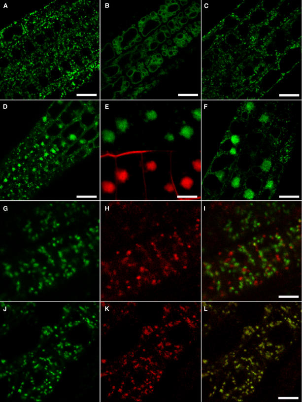
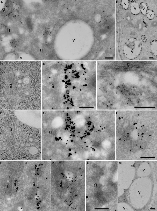
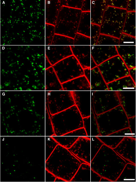
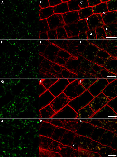
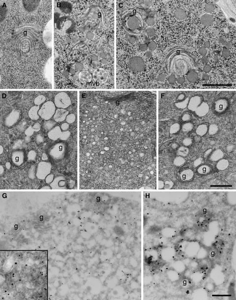
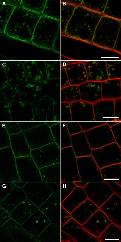
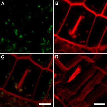
In eukaryotic cells, compartments of the highly dynamic endomembrane system are acidified to varying degrees by the activity of vacuolar H(+)-ATPases (V-ATPases). In the Arabidopsis thaliana genome, most V-ATPase subunits are encoded by small gene families, thus offering potential for a multitude of enzyme complexes with different kinetic properties and localizations. We have determined the subcellular localization of the three Arabidopsis isoforms of the membrane-integral V-ATPase subunit VHA-a. Colocalization experiments as well as immunogold labeling showed that VHA-a1 is preferentially found in the trans-Golgi network (TGN), the main sorting compartment of the secretory pathway. Uptake experiments with the endocytic tracer FM4-64 revealed rapid colocalization with VHA-a1, indicating that the TGN may act as an early endosomal compartment. Concanamycin A, a specific V-ATPase inhibitor, blocks the endocytic transport of FM4-64 to the tonoplast, causes the accumulation of FM4-64 together with newly synthesized plasma membrane proteins, and interferes with the formation of brefeldin A compartments. Furthermore, nascent cell plates are rapidly stained by FM4-64, indicating that endocytosed material is redirected into the secretory flow after reaching the TGN. Together, our results suggest the convergence of the early endocytic and secretory trafficking pathways in the TGN.


Figures

Figure 1.
Figure 1.
Subcellular Localization of VHA-a–GFP Fusion Proteins Expressed in Roots of Seedlings. (A) to (C) Cells in the root elongation zone expressing VHA-a1–GFP (A), VHA-a2–GFP (B), and VHA-a1a2–GFP (C). Bars = 25 μm. (D) to (F) Cells expressing VHA-a1–GFP ([D] and [E]) and VHA-a1a2–GFP (F) after treatment with BFA for 1 h. Costaining with FM4-64 is shown in (E). Bars = 25 μm ([D] and [F]) and 10 μm (E). (G) to (I) Cells coexpressing VHA-a1–GFP and the endosome marker ARA7-mRFP. Shown are the GFP channel (green [G]), the separately recorded mRFP channel (red [H]), and the overlay (I). Bar = 10 μm. (J) to (L) Cells coexpressing VHA-a1–GFP and the TGN marker SYP41-mRFP. Shown are the GFP channel (green [J]), the separately recorded mRFP channel (red [K]), and the overlay (L). Bar = 10 μm.
Figure 2.
Figure 2.
Immunogold Localization of VHA-a1–GFP and TGN Morphology. Immunogold labeling of ultrathin cryosections was performed with anti-GFP antibodies ([A], [D], [E], [G], and [I] to [L]) or anti–VHA-E antibodies ([B], [H], and [M]) and silver-enhanced Nanogold IgG. In (L), silver-enhanced Qdot525 IgG was used as a marker. (A) Overview of an immunogold-labeled cryosection of a root tip cortex cell expressing VHA-a1–GFP. Gold markers accumulate in the vicinity of Golgi stacks (g). (B) The anti–VHA-E antibody strongly labels vacuolar membranes (v) and, to a lesser extent, the TGN region (see [H]). (C) and (F) Electron micrographs of Golgi stacks and their associated TGNs in high-pressure frozen and freeze-substituted root tip cortex cells, shown at the same magnification as (D), (G), and (H). (D), (E), and (G) Golgi stacks and their associated TGNs after immunogold labeling of VHA-a1–GFP in root cortex cells. (H) Labeling of wild-type cells using anti–VHA-E antibodies results in gold labeling in the vicinity of Golgi stacks and on vacuolar membranes (see [B]). (I) The a1a2-GFP chimeric protein can be detected on the TGN. (J) and (K) N-ST-GFP is located at the trans-most Golgi cisternae (J) and often also in the TGN (K). Labeling of N-ST-GFP with the silver-enhanced Quantum dot marker (Qdot525) (J) resulted in a labeling pattern and label density similar to those of silver-enhanced Nanogold markers. (L) Control labeling on wild-type root cryosections using anti-GFP antibodies and silver-enhanced Nanogold was negligible. (M) Control labeling with anti–VHA-E antibodies on cryosections of a VHA-E1–deficient embryo (Strompen et al., 2005). There are only a few gold particles on vacuolar membranes. Bars = 0.25 μm.
Figure 3.
Figure 3.
Rapid Colocalization of VHA-a1–GFP with FM4-64. Cells in the root elongation zone expressing different GFP markers were stained with the endocytic tracer FM4-64: VHA-a1–GFP (A), N-ST-YFP (D), ARA7-GFP (G), and ARA6-GFP (J). Overlays of the separately recorded GFP (green) and FM4-64 (red) channels ([B], [E], [H], and [K]) are shown in (C), (F), (I), and (L). All images were taken after 6 min of FM4-64 uptake. Bars = 10 μm.
Figure 4.
Figure 4.
ConcA Blocks FM4-64 Transport to the Tonoplast. Cells in the root elongation zone expressing VHA-a1–GFP were stained with the endocytic tracer FM4-64. Shown are the GFP channel ([A], [D], [G], and [J]), the FM4-64 channel ([B], [E], [H], and [K]), and overlays of the separately recorded GFP (green) and FM4-64 (red) channels ([C], [F], [I], and [L]). Bars = 10 μm. (A) to (C) Untreated cells expressing VHA-a1–GFP 1 h after FM4-64 staining. Arrowheads mark FM4-64 staining separate from VHA-a1–GFP signal that might represent later endosomal compartments. (D) to (F) Untreated cells expressing VHA-a1–GFP 2 h after FM4-64 staining. (G) to (I) ConcA-treated seedling root cells expressing VHA-a1–GFP 2 h after FM4-64 staining. Note the absence of FM4-64 staining not associated with VHA-a1–GFP and of tonoplast staining. (J) to (L) ConcA-treated seedling root cells expressing VHA-a1–GFP 5 h after FM4-64 staining. The arrow indicates faint tonoplast staining.
Figure 5.
Figure 5.
ConcA Induces Changes of TGN and Golgi Stack Morphology and Interferes with BFA Action. (A) to (C) Electron micrographs of high-pressure frozen and freeze-substituted root tip cortex cells showing changes in Golgi and TGN morphology after ConcA treatment, such as the characteristic bending and swelling of the ends of cisternae (A) and the fragmentation of Golgi stacks and the accumulation of large vesicles ([B] and [C]). g, Golgi stack; mvb, multivesicular bodies. (D) to (F) Electron micrographs of chemically fixed root tip cortex cells showing vesicle agglomerations induced by ConcA (D), BFA (E), and ConcA followed by BFA (F). The content of ConcA-induced vesicles is lost after chemical fixation and dehydration at ambient temperature (cf. [C]). (G) and (H) Immunogold labeling of VHA-a1–GFP on cryosections of root tip cortex cells using anti-GFP antibodies and silver-enhanced Nanogold after ConcA treatment (G) and BFA treatment (H). BFA and ConcA compartments are labeled. The inset in (H) shows a 2.5× enlarged detail of a thicker cryosection with gold-labeled vesicles of different sizes. Bars = 0.5 μm.
Figure 6.
Figure 6.
ConcA Induces Intracellular BRI1-GFP Accumulation. Cells in the root elongation zone expressing BRI1-GFP were stained with the endocytic tracer FM4-64. Shown are the GFP channels ([A], [C], [E], and [G]) and overlays of the separately recorded GFP (green) and FM4-64 (red) channels ([B], [D], [F], and [H]). Bars = 10 μm. (A) and (B) Untreated cells expressing BRI1-GFP stained with FM4-64 for 10 min. (C) and (D) Cells expressing BRI1-GFP treated with ConcA for 2 h in the presence of FM4-64. (E) and (F) Cells expressing BRI1-GFP treated with CHX for 1 h in the presence of FM4-64. (G) and (H) Cells expressing BRI1-GFP treated with CHX for 1 h followed by treatment with ConcA + CHX for 1 h in the presence of FM4-64.
Figure 7.
Figure 7.
Nascent Cell Plates Are Rapidly Stained by FM4-64. (A) to (C) Dividing cells expressing VHA-a1–GFP were stained with the endocytic tracer FM4-64. Images were taken within 15 min of FM4-64 staining. Shown are the separately recorded GFP (A) and FM4-64 (B) channels as well as the overlay (C). (D) Three-dimensional reconstruction of an image stack derived from a Z scan shows that the cell plate is not connected to the surrounding plasma membrane. Bars = 5 μm.

References

    1. Ali, S., and Akazawa, T. (1986). Association of the H+-translocating ATPase in the Golgi membrane system from suspension-cultured cells of sycamore (Acer pseudoplantanus L.). Plant Physiol. 81 222–227. - PMC - PubMed
    1. Aniento, F., Gu, F., Parton, R.G., and Gruenberg, J. (1996). An endosomal beta COP is involved in the pH-dependent formation of transport vesicles destined for late endosomes. J. Cell Biol. 133 29–41. - PMC - PubMed
    1. Bassham, D.C., Sanderfoot, A.A., Kovaleva, V., Zheng, H., and Raikhel, N.V. (2000). AtVPS45 complex formation at the trans-Golgi network. Mol. Biol. Cell 11 2251–2265. - PMC - PubMed
    1. Bayer, M.J., Reese, C., Buhler, S., Peters, C., and Mayer, A. (2003). Vacuole membrane fusion: V0 functions after trans-SNARE pairing and is coupled to the Ca2+-releasing channel. J. Cell Biol. 162 211–222. - PMC - PubMed
    1. Belanger, K.D., and Quatrano, R.S. (2000). Membrane recycling occurs during asymmetric tip growth and cell plate formation in Fucus distichus zygotes. Protoplasma 212 24–37.

Publication types

MeSH terms